Apoptosis occurs during normal cellular development and involves dramatic changes in cellular structure. Disruption of apoptosis may contribute to cancer as well as other autoimmune diseases. Caspase family of cysteine proteases has been shown to play a key role in apoptosis. Caspase-8 is a 55 kDa cytosolic protein that is synthesized as an inactive pro-enzyme. Activation of caspase-8 involves a two-step proteolysis: the cleavage of caspase-8 to generate a 43 and a 12 kDa fragment which is further processed to 10 kDa. The p43 is then cleaved to yield p26 and the release of the active site containing p18.The Active/Cleaved Caspase-8 polyclonal antisera recognizes the large and small subunits of active/cleaved caspase-8. Whereas the antisera has a strong preference for active/cleaved caspase-8, in some cell or tissue systems or techniques the antisera may also recognize the proform of caspase-8 as well as intermediate caspase-8 cleavage fragments.
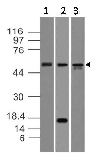
Fig-1:Western blot analysis of Caspase-8. Anti-Caspase-8 antibody (Clone: ABM14C1) was used at 2 µg/ml on Molt-4, Kato III and HepG2 lysates.
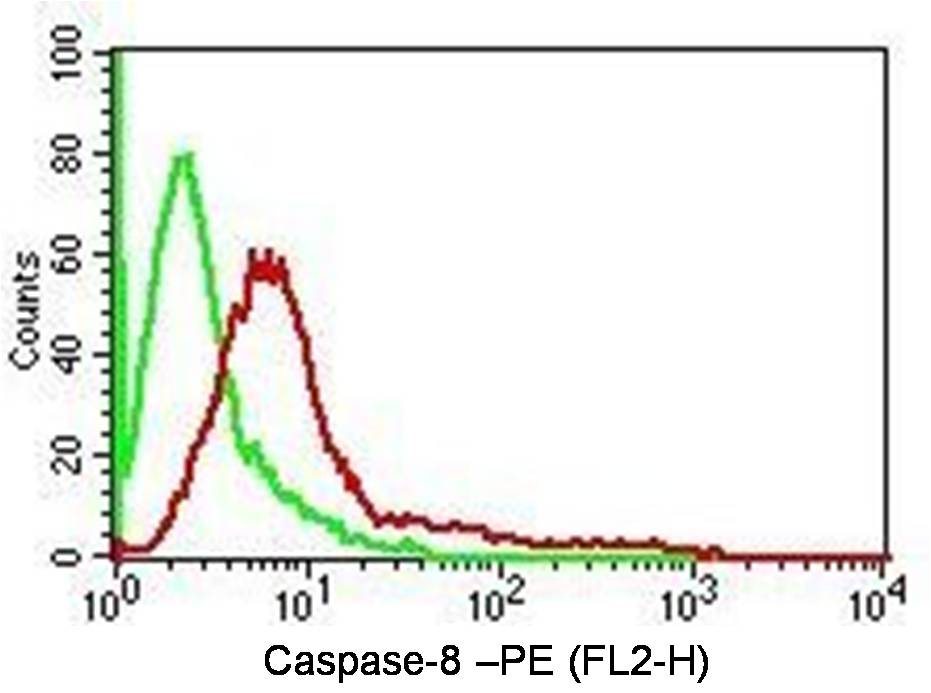
Fig-2: Intracellular Flow analysis of Caspase-8 antibody in Hela cells using 0.5 µg/ 10 6 cells of anti-Caspase-8 antibody (ABM14C1). Green represents isotype control, red represents anti-Caspase-8 antibody. Goat anti-mouse PE conjugate was used as secondary antibody. (Cells were fixed with 4% paraformaldehyde for 10 min and washed with PBS by centrifuging at 1100 for 5 min followed by permeabilization for 20 min and washed again as mentioned above. Then cell were incubated with primary ant
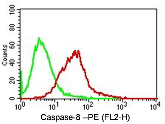
Fig-3: Intracellular Flow analysis of Caspase-8 antibody in Jurkat cells using 0.5 µg/ 10 6 cells of anti-Caspase-8 antibody (ABM14C1). Green represents isotype control, red represents anti-Caspase-8 antibody. Goat anti-mouse PE conjugate was used as secondary antibody. (Cells were fixed with 4% paraformaldehyde for 10 min and washed with PBS by centrifuging at 1100 for 5 min followed by permeabilization for 20 min and washed again as mentioned above. Then cell were incubated with primary a
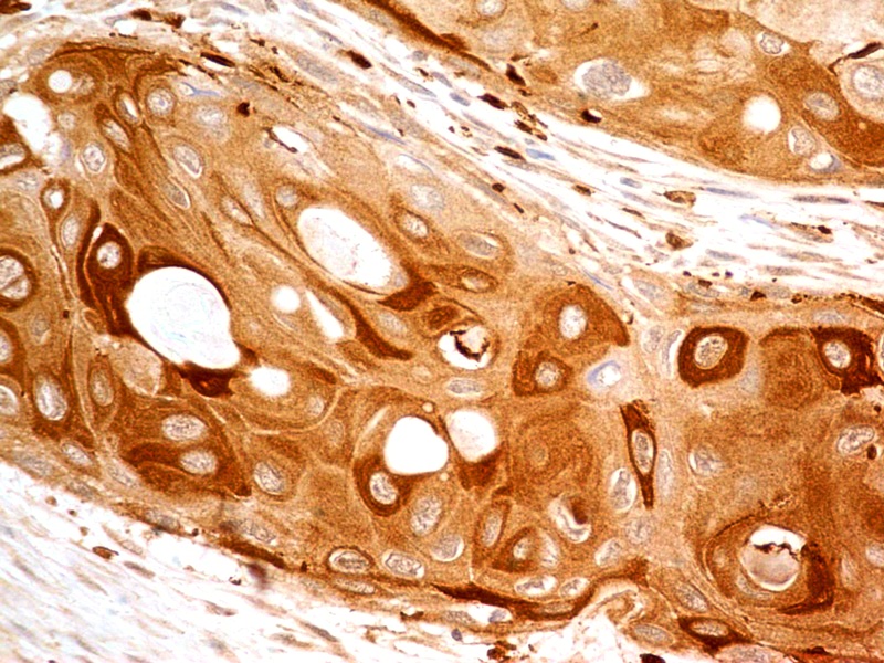
Fig-4: Immunohistochemical analysis of Caspase-8 in human Esophagus using anti-Caspase-8 antibody (Clone: ABM14C1) at 5 µg/ml.
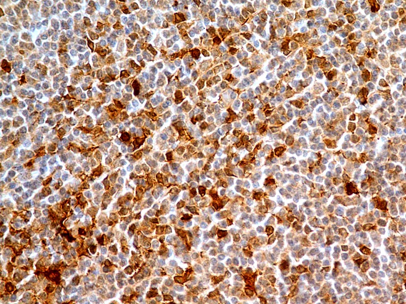
Fig-5: Immunohistochemical analysis of Caspase-8 in human Tonsil using anti-Caspase-8 antibody (Clone: ABM14C1) at 5 µg/ml.
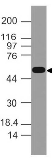
Figure-6: Western blot analysis of Caspase-8. Anti-Caspase-8 antibody (Clone: ABM14C1) was used at 4 µg/ml on EL-4 lysate.
* Mehrwertsteuer und Versandkosten nicht enthalten. Irrtümer und Preisänderungen vorbehalten