A partial length recombinant TLR2 protein (amino acids 180-420) was used as the immunogen for this antibody.
Alternative Synonym:
TLR2||TIL4
TLR2 (Toll-Like Receptors 2) is a member of the TLR (Toll-like receptor) family that plays a fundamental role in pathogen recognition and activation of innate immunity. TLR2 forms heterodimers with TLR1 and TLR6, which is the initial step in a cascade of events leading to significant innate immune responses, development of adaptive immunity to pathogens and protection from immune sequelae related to infection with these pathogens. TLR2 also interacts with a large number of non-TLR molecules, allowing for recognition of a great number and variety of PAMPs (pathogen-associated molecular patterns). TLR2 expression has been detected in immune cells, endothelial, and epithelial cells.
Western blot analysis: 2-4 µg/ml, FACS analysis: 0.5 µg/10 6 cells, Immunohistochemical analysis: 5 µg/ml
Fig-6: Immunohistochemical analysis of TLR2 in human Colon tissue using TLR2 antibody (Clone: ABM3A87) at 10 µg/ml.
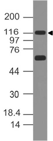
Fig-1: Western blot analysis of TLR2. Anti- TLR2 antibody (Clone: ABM3A87) was used at 2 µg/ml on mouse embryonic liver lysate.
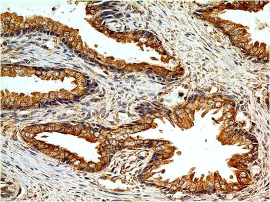
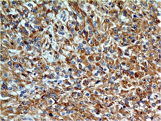
Fig-2 : Immunohistochemical analysis of TLR2 in human prostate tissue using TLR2 antibody (Clone: ABM3A87) at 5 µg/ml.
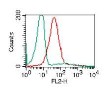
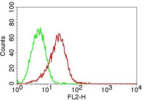
Fig-3: Intracellular flow analysis of TLR2 in PBMC (Monocytes) using 0.5 µg/10 6 cells of TLR2 antibody (Clone: ABM3A87). Green represents isotype control, red represents anti-TLR2 antibody. Goat anti-Mouse PE conjugate was used as secondary.
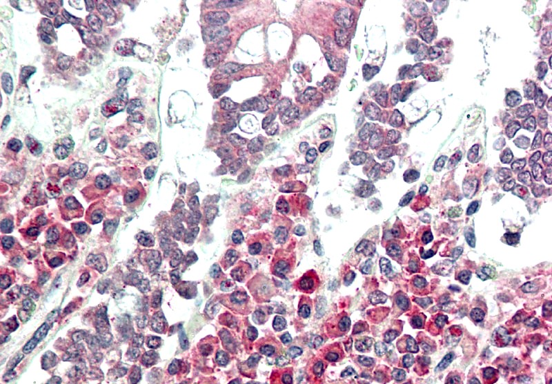
Fig-5: Immunohistochemical analysis of TLR2 in Renal Cell Carcinoma using TLR2 antibody (Clone: ABM3A87) at 5 µg/ml.
Fig-4: Intracellular flow analysis of TLR2 in THP-1 cells using 0.5 µg/10 6 cells of TLR2 antibody (Clone: ABM3A87). Green represents isotype control, red represents anti-TLR2 antibody. Goat anti-Mouse PE conjugate was used as secondary antibody.
* Mehrwertsteuer und Versandkosten nicht enthalten. Irrtümer und Preisänderungen vorbehalten