
Monoclonal Antibody to CD69 (Clone: ABM39A4) BSA Free
Artikelnummer:
ABI-10-4110-BSA-FREE
- Bilder (5)
| Artikelname: | Monoclonal Antibody to CD69 (Clone: ABM39A4) BSA Free |
| Artikelnummer: | ABI-10-4110-BSA-FREE |
| Hersteller Artikelnummer: | 10-4110-BSA-Free |
| Alternativnummer: | ABI-10-4110-BSA-FREE-100UG |
| Hersteller: | Abeomics |
| Wirt: | Mouse |
| Kategorie: | Antikörper |
| Applikation: | FACS, IHC, WB |
| Spezies Reaktivität: | Human |
| Immunogen: | Full length recombinant CD69 protein was used as the immunogen for this antibody. |
| Alternative Synonym: | CD69||CLEC2C |
| CD69 is a type II transmembrane protein of the C-type lectin family and its gene is located within the natural killer gene complex. In healthy subjects, CD69 is not detected in peripheral blood lymphocytes, but is expressed on small subsets of T and B cells in peripheral lymphoid tissues. CD69 gene maps at human chromosome 12, and behaves as an early activation gene that contains responsive elements for the transcription factors NF-kappaB, ERG-1 and AP-1. This protein acts as a leukocyte activation marker, and is involved in the activation of different leukocyte subsets as well as in the pathogenesis of chronic inflammation. |
| Application Verdünnung: | Western blot analysis: 2-4 µg/ml, Immunohistochemical analysis: 5 µg/ml |
|
|
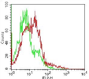
Fig-1: Cell surface FLOW analysis of hCD69 (10-4110) in PBMC treated 3 days with PMA + Ionomycin (Lymphocytes gated) using 0.5 µgantibody per 10 6 cells. Green represents PBMC W/O treatment, red represents PBMC with treatment. Goat anti-mouse PE conjugated secondary antibody was used (ABEOMICS). |
|
|
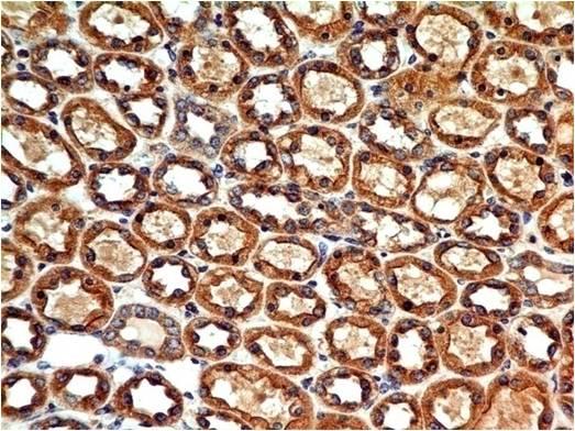
Fig-2 : Immunohistochemical analysis of CD69 in human kidney tissue using CD69 antibody (Clone: ABM39A4) at 5 µg/ml. |
|
|
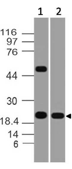
Figure-4: Western blot analysis of CD69. Anti- CD69 antibody (Clone: ABM39A4) was used at 4 µg/ml on (1)Daudi and (2) h Lung lysates. |
|
|
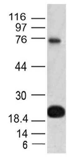
Figure-5: Western blot analysis of CD69. Anti- CD69 antibody (Clone: ABM39A4) was used at 2 µg/ml on U937 lysate. |
|
|
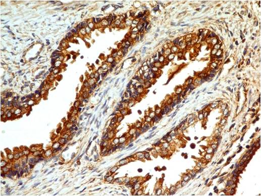
Figure-3: Immunohistochemical analysis of CD69 in human Prostate tissue using CD69 antibody (Clone: ABM39A4) at 5 µg/ml. |
Produktgarantie und fachkundiger Support
