Western blot analysis: 4-6 µg/ml, Flowcytometric analysis: 0.5-1 µg/10 6 cells
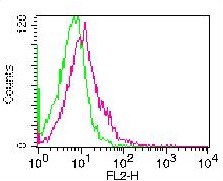
Figure 3: Cell surface staying of PHA stimulated PBMC. Green represents un-stimulated PBMC, red represents stimulated PBMC. 0.5 ug antibody was used. Goat anti-mouse PE was used as secondary antibody.
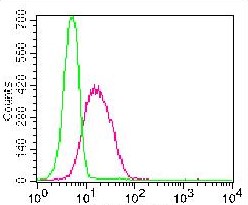
Figure:1- Cell surface staining of U937 cells. Green: Isotope control, Red: anti-Vista (10-7617) antibody. 0.5 µg antibody was used. Goat anti-mouse PE was used as secondary antibody.
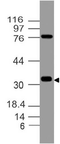
Figure:2- Western blot analysis of Vista. Anti-Vista antibody (Clone: ABM5C53) was tested at 4 µg/ml on h Testis Lysate.
* Mehrwertsteuer und Versandkosten nicht enthalten. Irrtümer und Preisänderungen vorbehalten