
Monoclonal antibody to SLC31A1 (Clone: ABM5D89)
Artikelnummer:
ABI-10-7652-25
- Bilder (8)
| Artikelname: | Monoclonal antibody to SLC31A1 (Clone: ABM5D89) |
| Artikelnummer: | ABI-10-7652-25 |
| Hersteller Artikelnummer: | 10-7652-25 |
| Alternativnummer: | ABI-10-7652-25-25UG |
| Hersteller: | Abeomics |
| Wirt: | Mouse |
| Kategorie: | Antikörper |
| Applikation: | IHC, WB |
| Spezies Reaktivität: | Human |
| Immunogen: | Full length recombinant SLC31A1 protein was used as the immunogen for this antibody. |
| Alternative Synonym: | Copper transporter 1, Solute carrier family 31 member 1, COPT1, CTR1 |
| Application Verdünnung: | Recommended dilutions: Western blot analysis: 1-2 µg/ml, Immunohistochemical analysis: 5 µg/ml. However, this need to be optimized based on the research applications. |
|
|
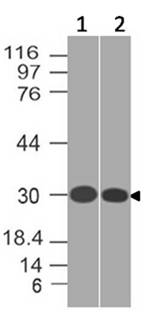
Figure-1: Western blot analysis of SLC31A1. Anti-SLC31A1 antibody (Clone: ABM5D89) was tested at 1 µg/ml on (1) T98G and (2) THP1 lysates. |
|
|
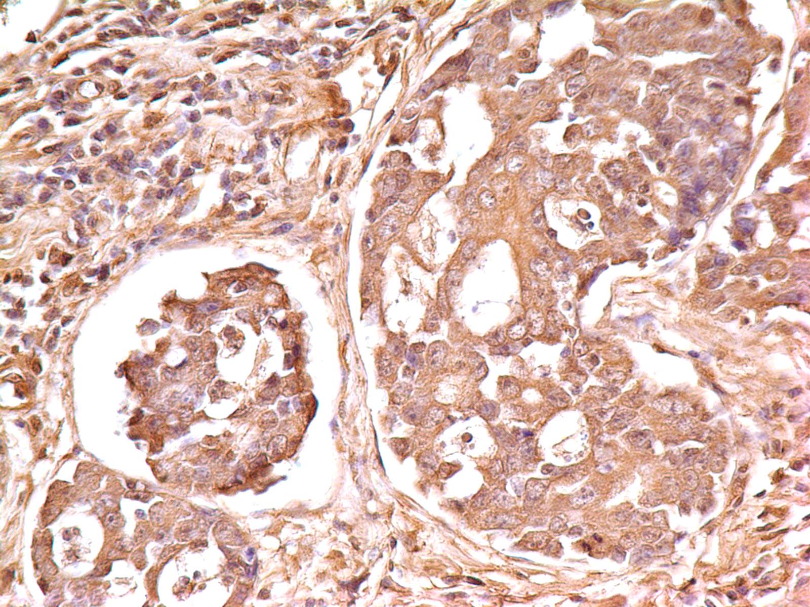
Figure-2: Immunohistochemical analysis of SLC31A1 in Adenocarcinoma cell of human Stomach using Anti-SLC31A1 antibody (Clone: ABM5D89) at 5 µg/ml. |
|
|
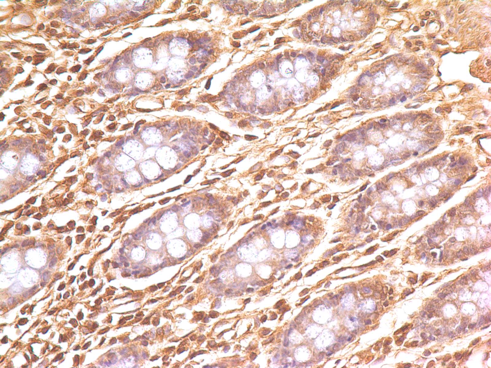
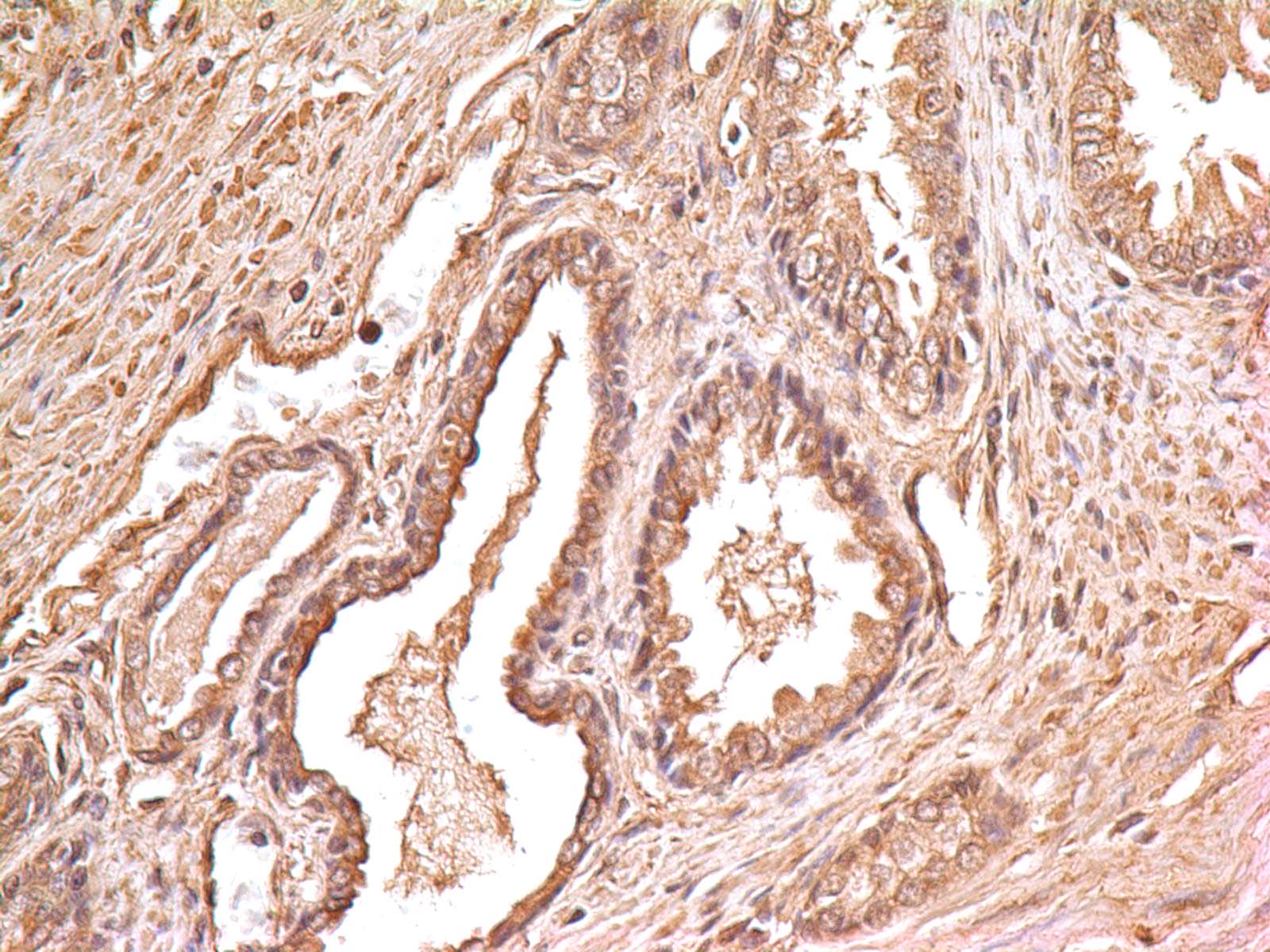
Figure-3: Immunohistochemical analysis of SLC31A1 in human Colon tissue using Anti-SLC31A1 antibody (Clone: ABM5D89) at 5 µg/ml. |
|
|
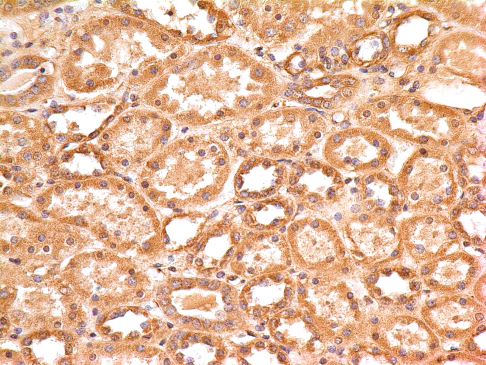
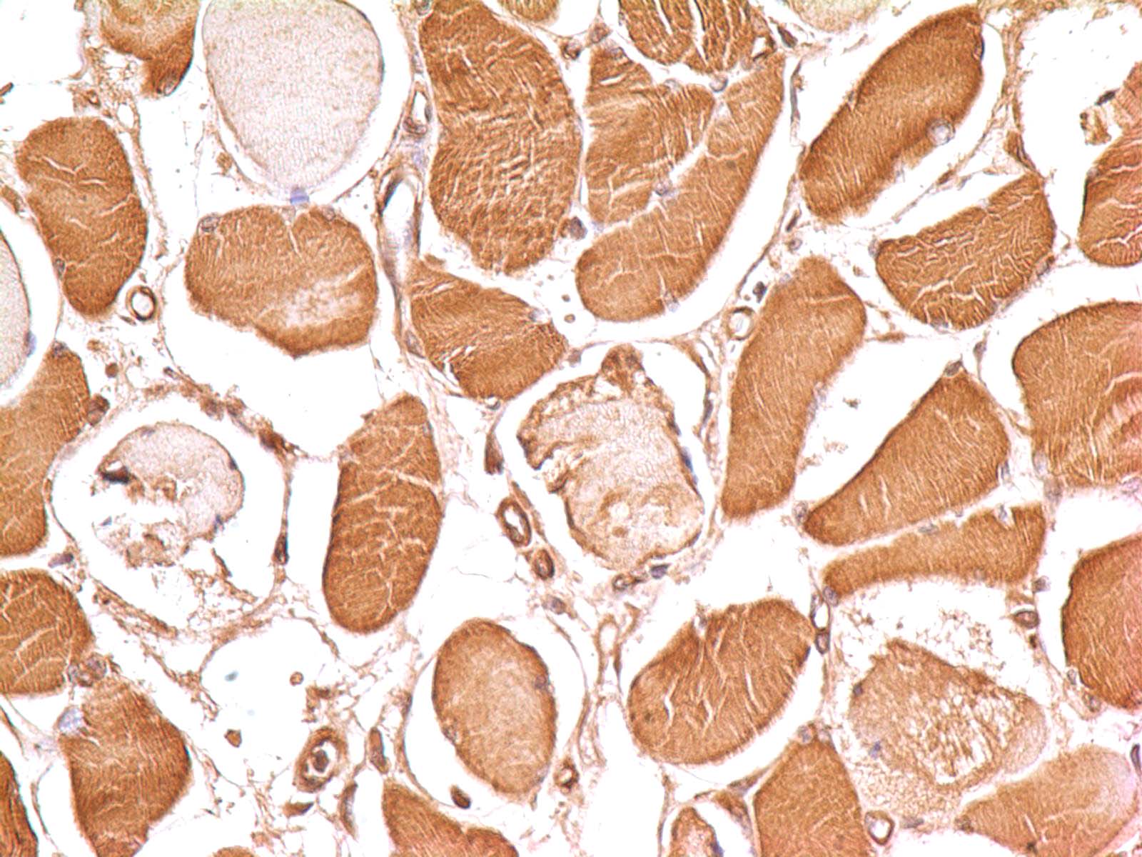
Figure-4: Immunohistochemical analysis of SLC31A1 in human Kidney tissue using Anti-SLC31A1 antibody (Clone: ABM5D89) at 5 µg/ml. |
|
|
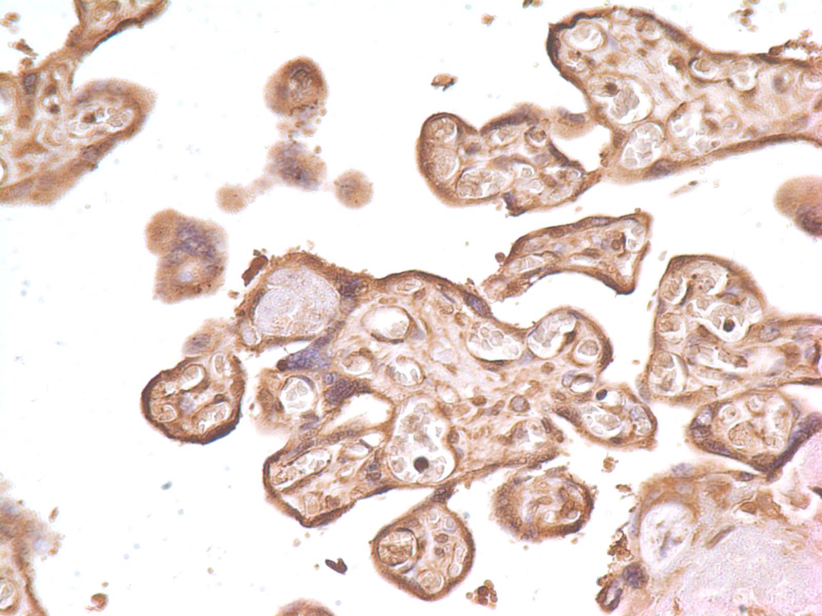
Figure-5: Immunohistochemical analysis of SLC31A1 in human Placenta tissue using Anti-SLC31A1 antibody (Clone: ABM5D89) at 5 µg/ml. |
|
|
Figure-6: Immunohistochemical analysis of SLC31A1 in human Prostate tissue using Anti-SLC31A1 antibody (Clone: ABM5D89) at 5 µg/ml. |
|
|
Figure-7: Immunohistochemical analysis of SLC31A1 in human Skin tissue using Anti-SLC31A1 antibody (Clone: ABM5D89) at 5 µg/ml. |
|
|
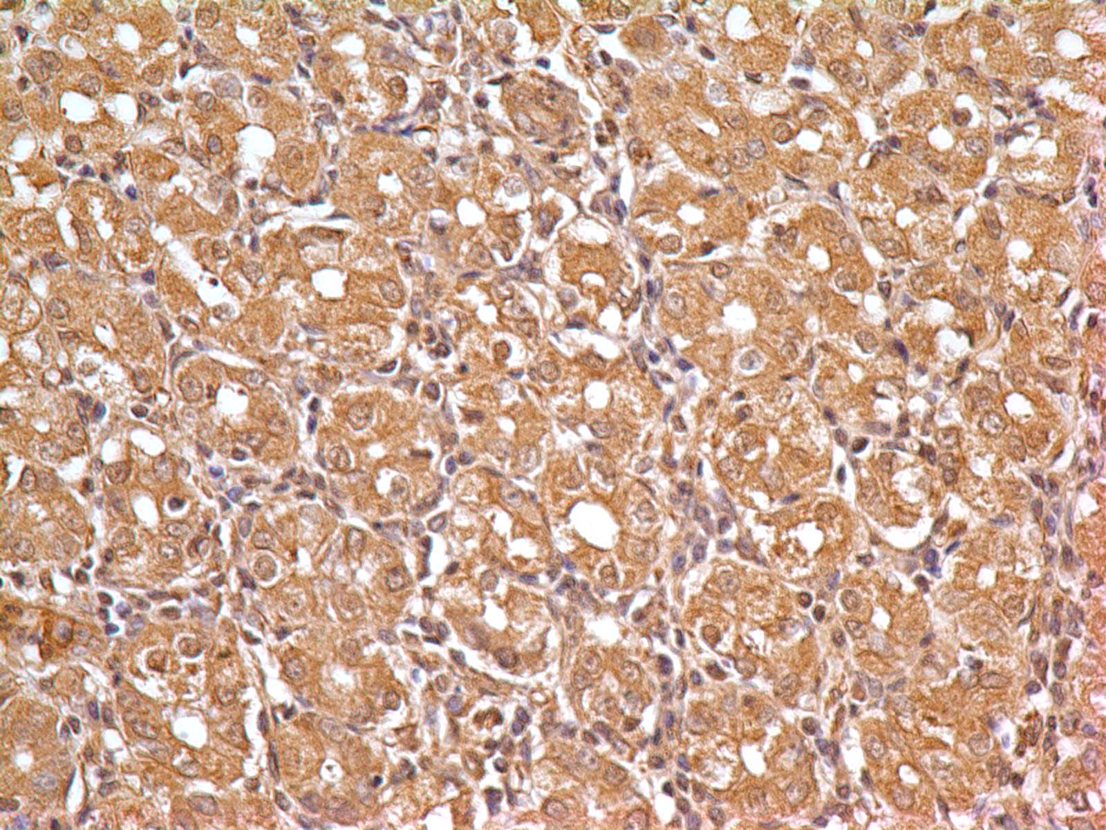
Figure-8: Immunohistochemical analysis of SLC31A1 in human Stomach tissue using Anti-SLC31A1 antibody (Clone: ABM5D89) at 5 µg/ml. |
Produktgarantie und fachkundiger Support
