Activated human peripheral blood mononuclear cells
Alternative Synonym:
HLA-DRB1||HLA-DRB2
This MAb reacts with a 28kDa chain of HLA-DRB1 antigen, a member of MHC class II molecules. It does not cross react with HLA-DP and HLA-DQ. The L243 antibody recognizes a different epitope than the LN3 monoclonal antibody, and these antibodies do not cross-block binding to each others respective epitopes. HLA-DR is a heterodimeric cell surface glycoprotein comprised of a 36kDa alpha (heavy) chain and a 28kDa beta (light) chain. It is expressed on B-cells, activated T-cells, monocytes/macrophages, dendritic cells and other non-professional APCs. In conjunction with the CD3/TCR complex and CD4 molecules, HLA-DR is critical for efficient peptide presentation to CD4+ T cells. It is an excellent histiocytic marker in paraffin sections producing intense staining. True histiocytic neoplasms are similarly positive. HLA-DR antigens also occur on a variety of epithelial cells and their corresponding neoplastic counterparts. Loss of HLA-DR expression is related to tumor microenvironment and predicts adverse outcome in diffuse large B-cell lymphoma.
Flow Cytometry (1-2ug/million cells), Immunofluorescence (1-2ug/ml), Western Blot (1-2ug/ml), Immunohistochemistry (Formalin-fixed) (1-2ug/ml for 30 minutes at RT)(Staining of formalin-fixed tissues requires heating tissue sections in 10mM Tris with 1mM E
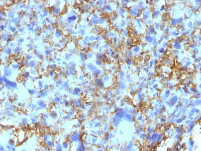
Formalin-fixed, paraffin-embedded human Histiocytoma stained with HLA-DRB Monoclonal Antibody (LN-3).
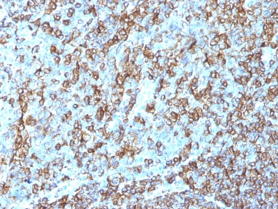
Formalin-fixed, paraffin-embedded human Tonsil stained with HLA-DRB Monoclonal Antibody (LN-3).
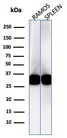
Western Blot Analysis of Ramos cell and human spleen lysate using HLA-DR Mouse Monoclonal Antibody (LN-3).
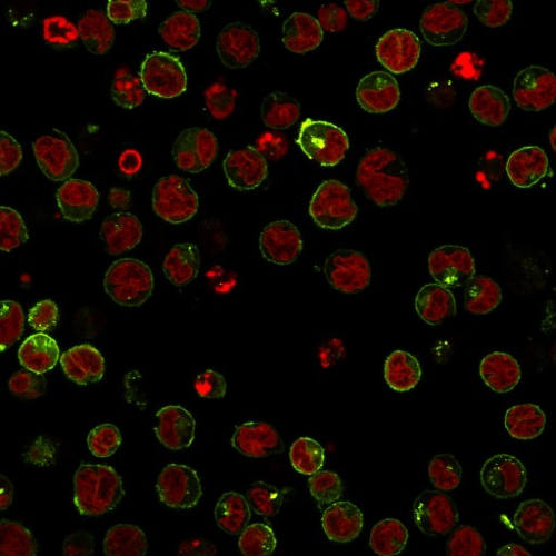
Immunofluorescent staining of Raji cells. HLA-DR Mouse Monoclonal Antibody (LN-3) labeled with CF594 (green) The nuclear counterstain is RedDot (red).
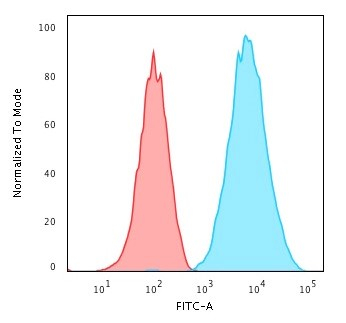
Flow Cytometric Analysis of Raji cells. HLA-DR Mouse Monoclonal Antibody (LN-3) followed by goat anti-mouse IgG-CF488 (blue), isotype control (red).
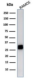
Western Blot Analysis of Ramos cell lysate using HLA-DR Mouse Monoclonal Antibody (LN-3).
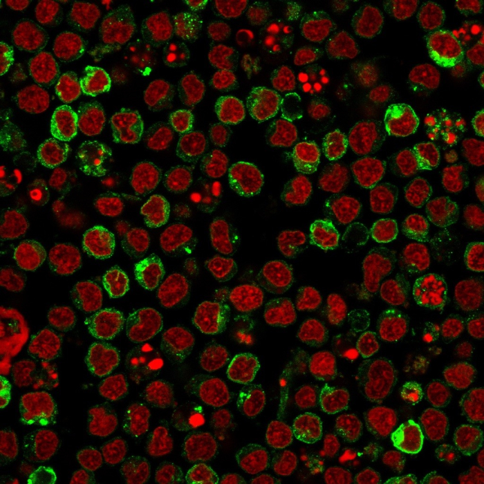
Immunofluorescence staining of Ramos cells. HLA-DR Mouse Monoclonal Antibody (LN-3) followed by goat anti-mouse IgG-CF488 (green). Nuclei counterstain is RedDot.
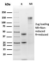
SDS-PAGE Analysis Purified HLA-DR Mouse Monoclonal Antibody (LN-3). Confirmation of Integrity and Purity of Antibody.
* Mehrwertsteuer und Versandkosten nicht enthalten. Irrtümer und Preisänderungen vorbehalten