
Anti-MEF2C Recombinant Antibody, IgG1, Clone: [CL0368], Mouse, Monoclonal
Artikelnummer:
ATA-AMAB90727R
- Bilder (9)

| Artikelname: | Anti-MEF2C Recombinant Antibody, IgG1, Clone: [CL0368], Mouse, Monoclonal |
| Artikelnummer: | ATA-AMAB90727R |
| Hersteller Artikelnummer: | AMAb90727R |
| Alternativnummer: | ATA-AMAB90727R-100,ATA-AMAB90727R-25 |
| Hersteller: | Atlas Antibodies |
| Wirt: | Mouse |
| Kategorie: | Antikörper |
| Applikation: | ICC, IHC, WB |
| Spezies Reaktivität: | Human |
| Recombinant Mouse Monoclonal Anti-MEF2C Antibody against Human myocyte enhancer factor 2C. Validated for Immunofluorescence, Immunohistochemistry and Western Blot |
|
|
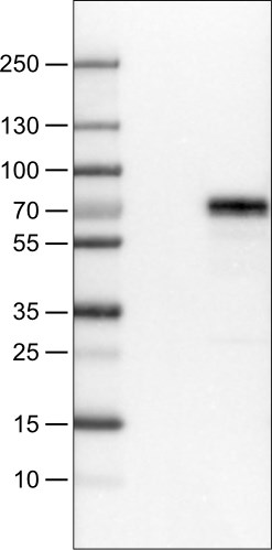
Lane 1: Marker [kDa] Lane 2: Negative control (vector only transfected HEK293T lysate) Lane 3: MEF2C Over-expression Lysate (Co-expressed with a C-terminal myc-DDK tag (~3.1 kDa) in mammalian HEK293T cells, LY419349) |
|
|
Immunohistochemical staining of human cerebellum shows strong nuclear positivity in Purkinje cells. |
|
|
Immunohistochemical staining of human cerebral cortex shows strong immunoreactivity in neuronal cells. |
|
|
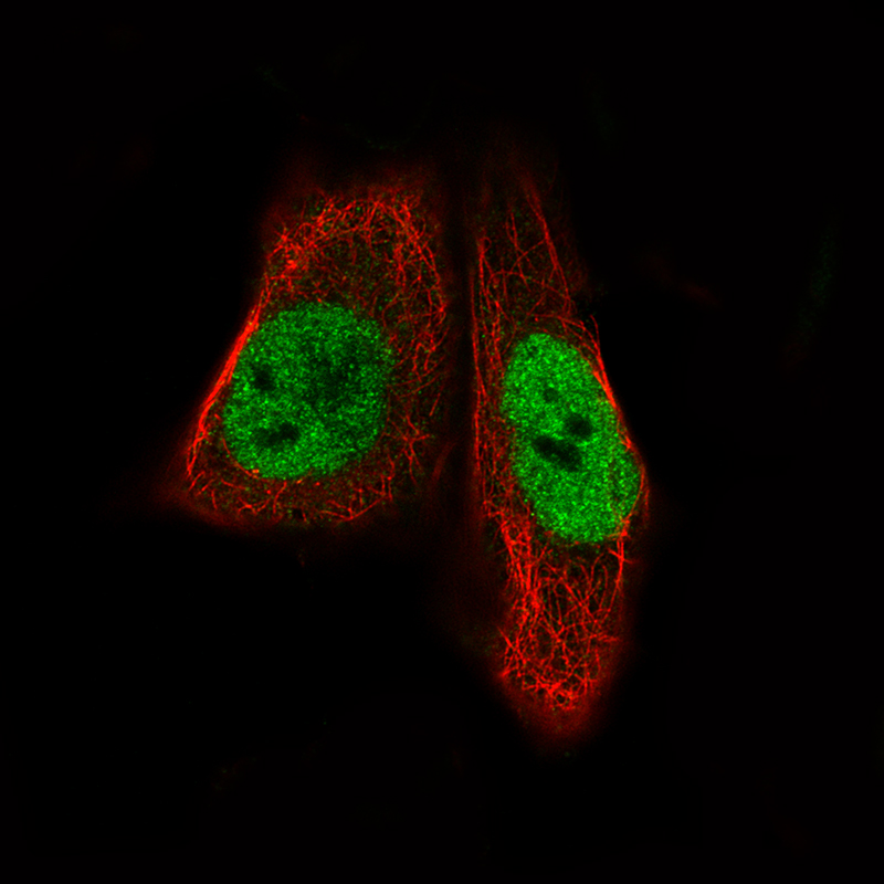
Immunofluorescence staining of RH-30 cells using the Anti-MEF2C monoclonal antibody, showing specific staining in the nucleoplasm in green. Microtubule- and nuclear probes are visualized in red and blue, respectively (where available). |
|
|
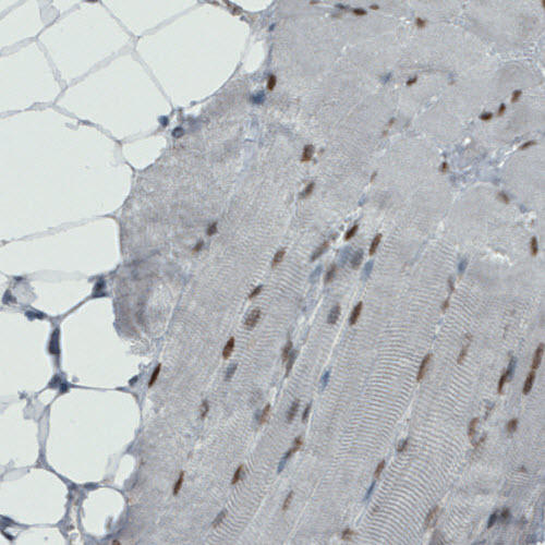
Immunohistochemical staining of human skeletal muscle shows moderate nuclear positivity in muscle fibers. |
|
|
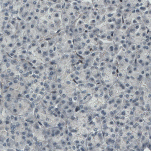
Immunohistochemical staining of human pancreas shows absense of staining (negative control). |
|
|
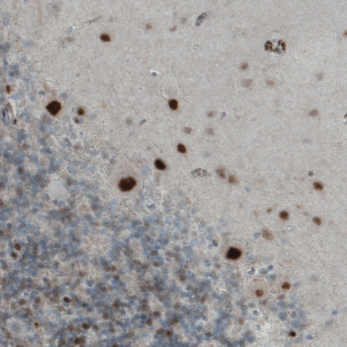
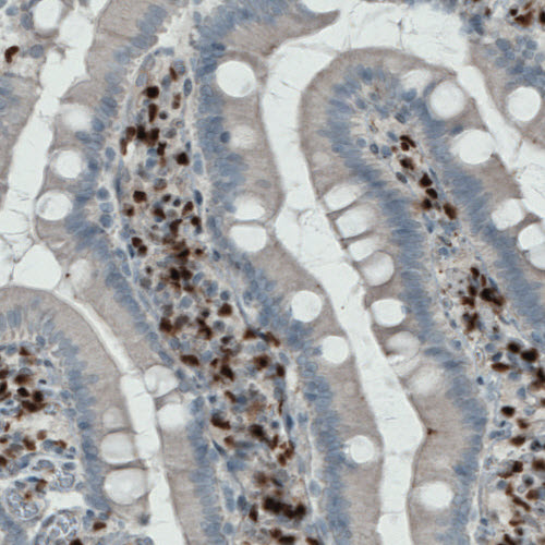
Immunohistochemical staining of human duodenum shows strong nuclear immunoreactivity in a subset of lymphoid cells. |
|
|
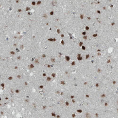
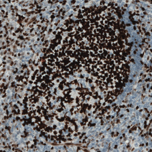
Immunohistochemical staining of human tonsil shows strong nuclear immunoreactivity in a subset of lymphoid cells. |
|
|
Western blot analysis in WM115 cells transfected with control siRNA, target specific siRNA probe 1 and 2, using Anti-MEF2C antibody. Remaining relative intensity is presented. Loading control: Anti-GAPDH. |
Produktgarantie und fachkundiger Support
