
Anti-TUFM Recombinant Antibody, IgG1, Clone: [CL2242], Mouse, Monoclonal
Artikelnummer:
ATA-AMAB90964R
- Bilder (12)
| Artikelname: | Anti-TUFM Recombinant Antibody, IgG1, Clone: [CL2242], Mouse, Monoclonal |
| Artikelnummer: | ATA-AMAB90964R |
| Hersteller Artikelnummer: | AMAb90964R |
| Alternativnummer: | ATA-AMAB90964R-100, ATA-AMAB90964R-25 |
| Hersteller: | Atlas Antibodies |
| Wirt: | Mouse |
| Kategorie: | Antikörper |
| Applikation: | ICC, IHC, WB |
| Spezies Reaktivität: | Human |
| Alternative Synonym: | EF-TuMT, EFTu |
| Recombinant Mouse Monoclonal Anti-TUFM Antibody against Human Tu translation elongation factor, mitochondrial. Validated for Immunofluorescence, Immunohistochemistry and Western Blot |
|
|
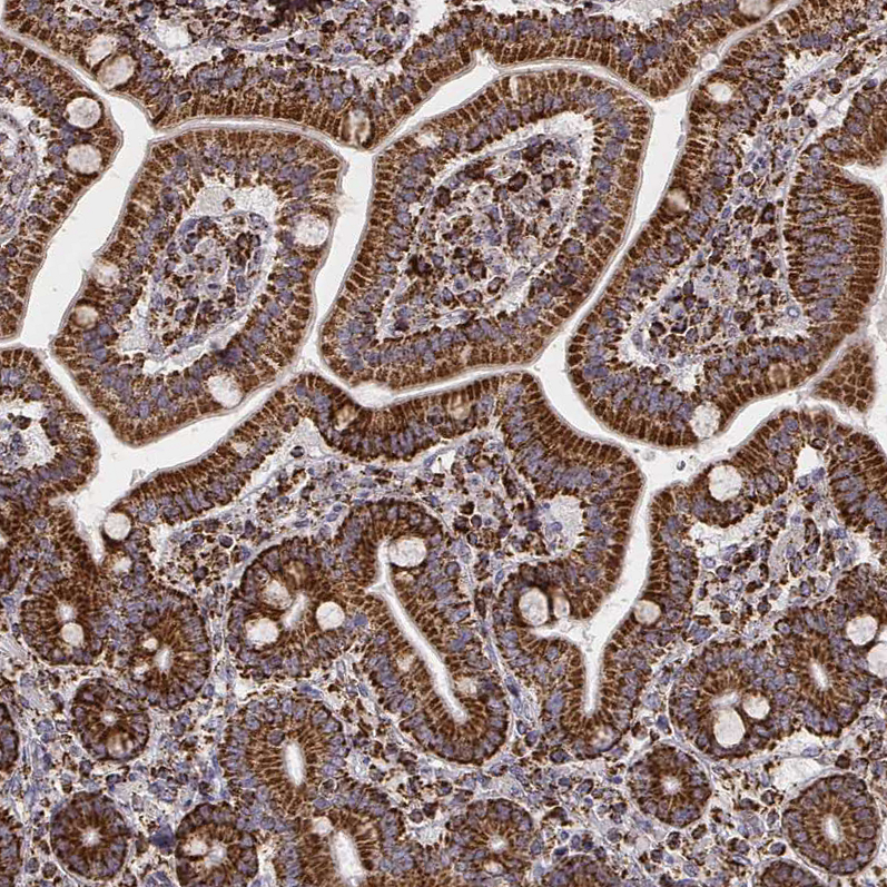
Immunohistochemical staining of human duodenum shows strong granular cytoplasmic positivity in glandular cells. |
|
|
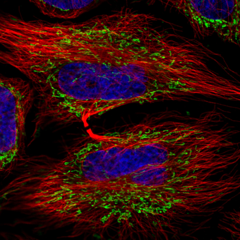
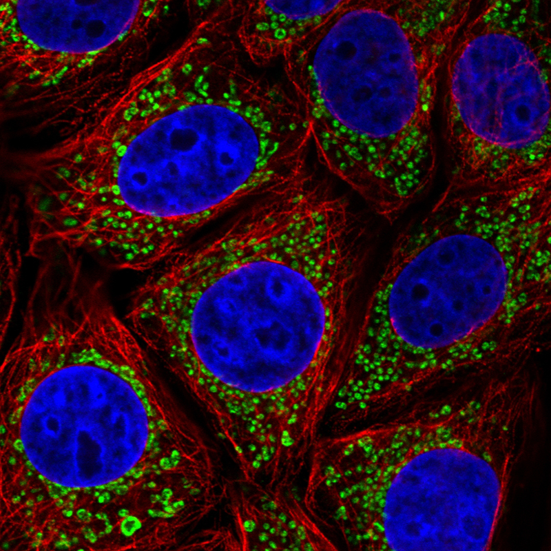
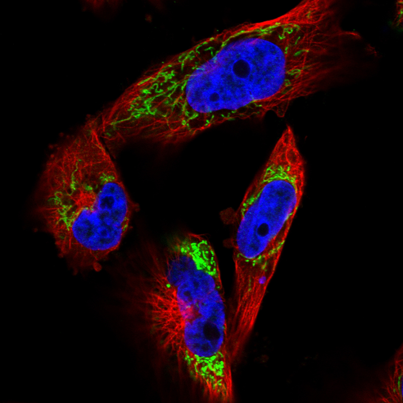
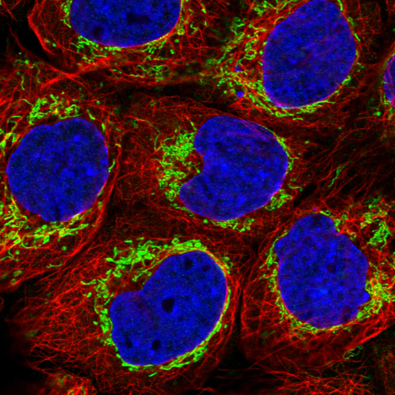
Immunofluorescence staining in HeLa cell line with Anti-TUFM monoclonal antibody, showing distinct mitochondrial staining in green. Microtubule- and nuclear probes are visualized in red and blue respectively (where available). |
|
|
Immunofluorescence staining in U2OS cell line with Anti-TUFM monoclonal antibody, showing distinct mitochondrial staining in green. Microtubule- and nuclear probes are visualized in red and blue respectively (where available). |
|
|
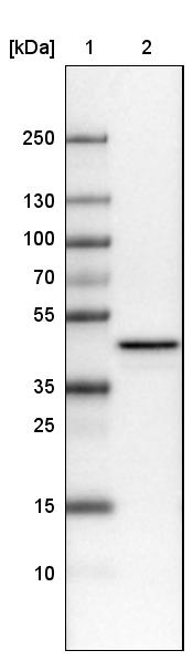
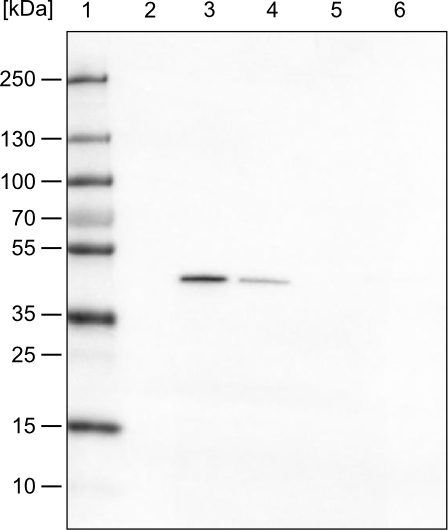
Lane 1: Marker [kDa] Lane 2: Human cell line U-251 MG |
|
|
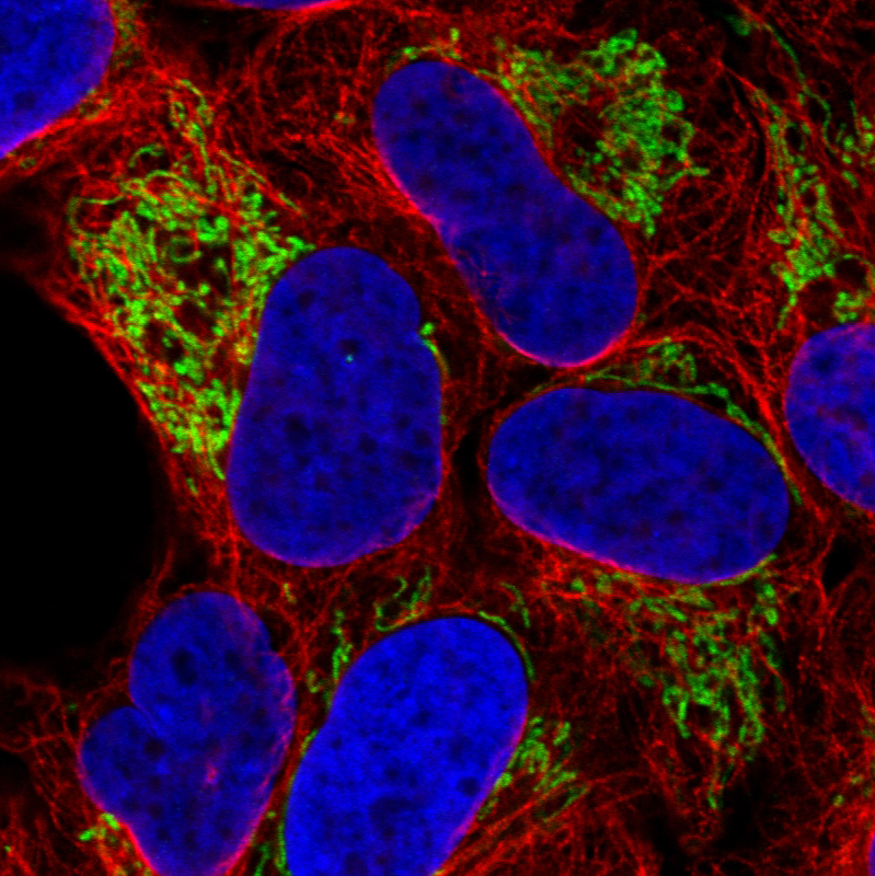
Immunofluorescence staining in MCF7 cell line with Anti-TUFM monoclonal antibody, showing distinct mitochondrial staining in green. Microtubule- and nuclear probes are visualized in red and blue respectively (where available). |
|
|
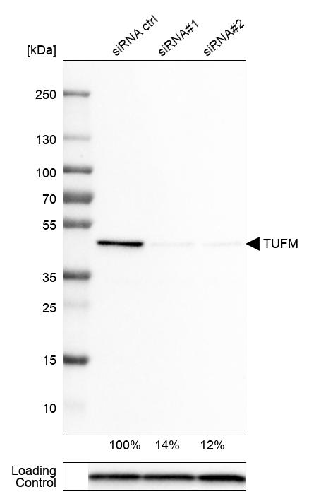
Western blot analysis in U-251MG cells transfected with control siRNA, target specific siRNA probe 1 and 2, using Anti-TUFM antibody. Remaining relative intensity is presented. Loading control: Anti-PPIB. |
|
|
Immunofluorescence staining in U251 cell line with Anti-TUFM monoclonal antibody, showing distinct mitochondrial staining in green. Microtubule- and nuclear probes are visualized in red and blue respectively (where available). |
|
|
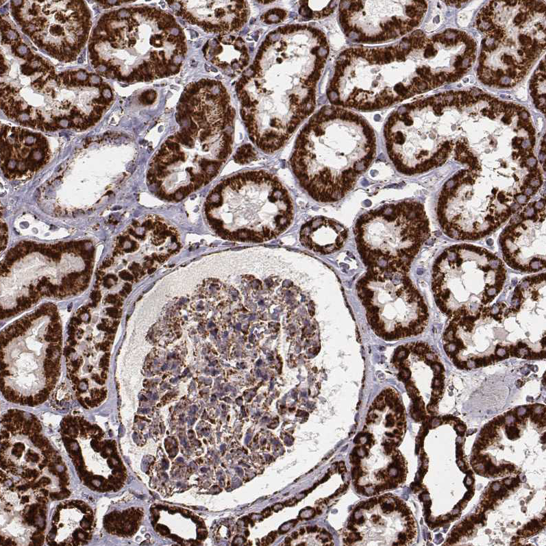
Immunohistochemical staining of human kidney shows strong granular cytoplasmic positivity in cells in tubules and moderate staining in glomerular cells. |
|
|
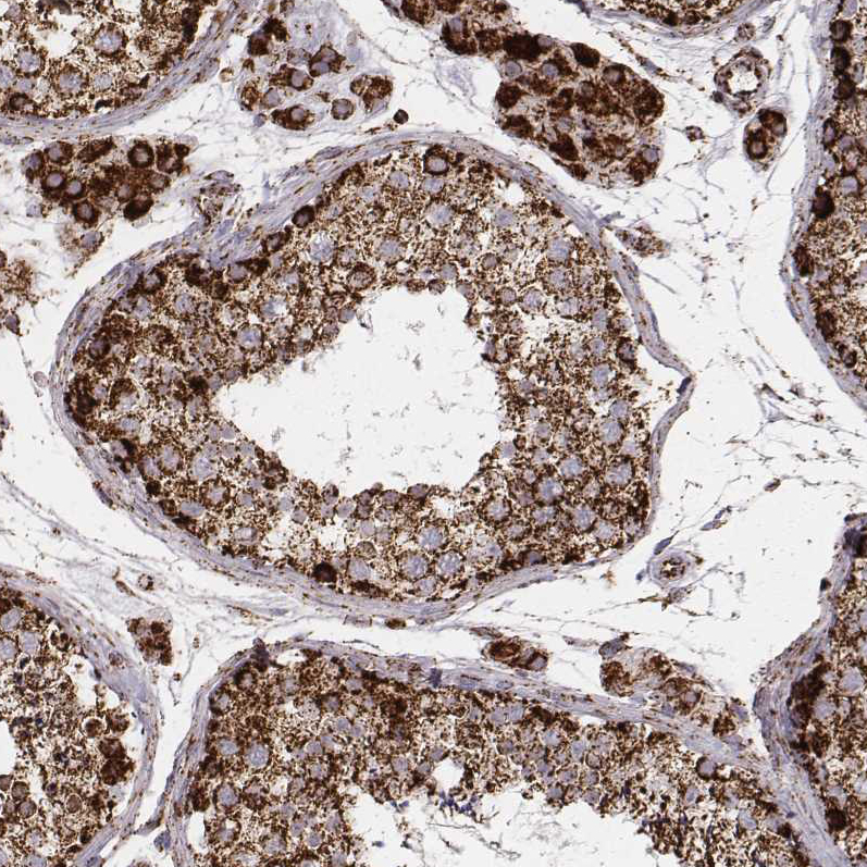
Immunohistochemical staining of human testis shows strong granular cytoplasmic positivity in cells in seminiferous ducts and in Leydig cells. |
|
|
Immunofluorescence staining in A431 cell line with Anti-TUFM monoclonal antibody, showing distinct mitochondrial staining in green. Microtubule- and nuclear probes are visualized in red and blue respectively (where available). |
|
|
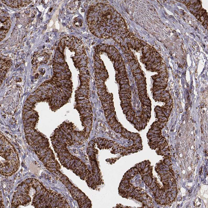
Immunohistochemical staining of human fallopian tube shows strong granular cytoplasmic positivity in glandular cells. |
|
|
Lane 1: Marker [kDa] Lane 2: Human cell line HeLa cytoplasmic fraction Lane 3: Human cell line HeLa membrane fraction Lane 4: Human cell line HeLa nuclear fraction Lane 5: Human cell line HeLa chromatin fraction Lane 6: Human cell line HeLa cytoskeletal fraction |
Produktgarantie und fachkundiger Support
