
Anti-ABCD3 Recombinant Antibody, IgG1, Clone: [CL2524], Mouse, Monoclonal
Artikelnummer:
ATA-AMAB90995R
- Bilder (12)
| Artikelname: | Anti-ABCD3 Recombinant Antibody, IgG1, Clone: [CL2524], Mouse, Monoclonal |
| Artikelnummer: | ATA-AMAB90995R |
| Hersteller Artikelnummer: | AMAb90995R |
| Alternativnummer: | ATA-AMAB90995R-100, ATA-AMAB90995R-25 |
| Hersteller: | Atlas Antibodies |
| Wirt: | Mouse |
| Kategorie: | Antikörper |
| Applikation: | ICC, IHC |
| Spezies Reaktivität: | Human |
| Alternative Synonym: | PMP70, PXMP1, ZWS2 |
| Recombinant Mouse Monoclonal Anti-ABCD3 Antibody against Human Atp binding cassette subfamily d member 3. Validated for Immunofluorescence and Immunohistochemistry |
|
|
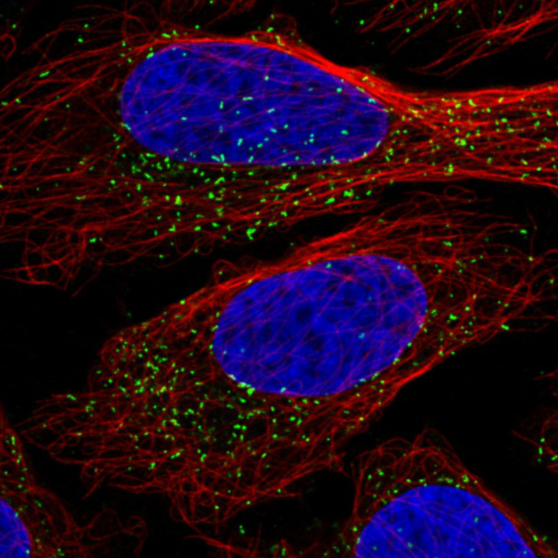
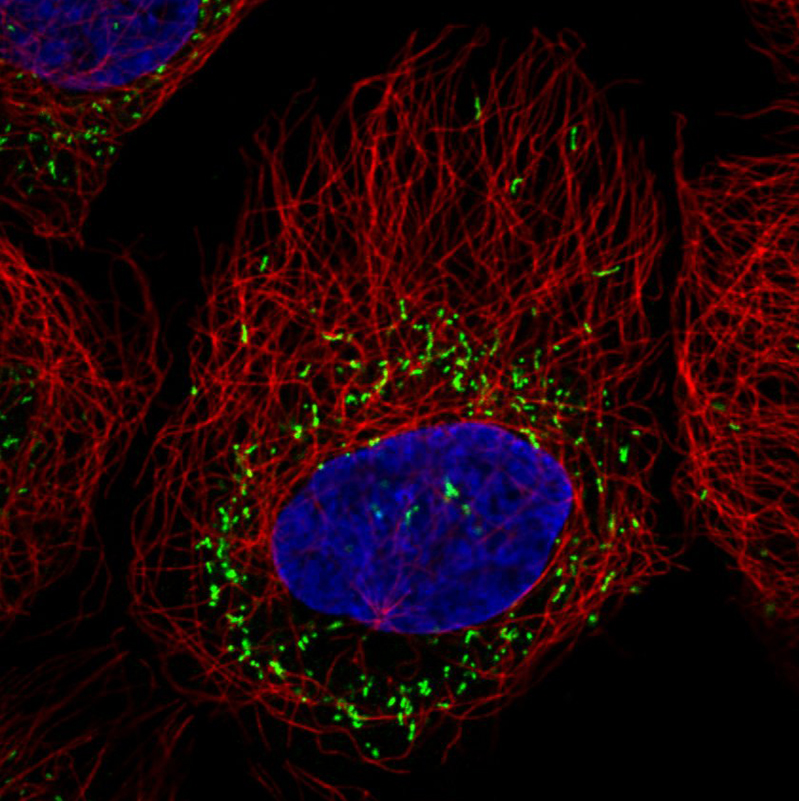
Immunofluorescence staining in U2OS cell line with Anti-ABCD3 monoclonal antibody, showing specific staining of peroxisomes in green. Microtubule- and nuclear probes are visualized in red and blue respectively (where available). |
|
|
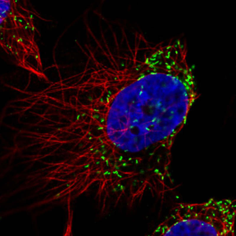
Immunofluorescence staining in U251 cell line with Anti-ABCD3 monoclonal antibody, showing specific staining of peroxisomes in green. Microtubule- and nuclear probes are visualized in red and blue respectively (where available). |
|
|
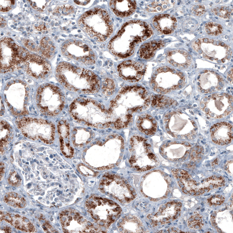
Immunohistochemical staining of human kidney shows moderate granular cytoplasmic positivity in cells in tubules. |
|
|
Immunohistochemical staining of human rectum shows moderate granular cytoplasmic positivity in glandular cells. |
|
|
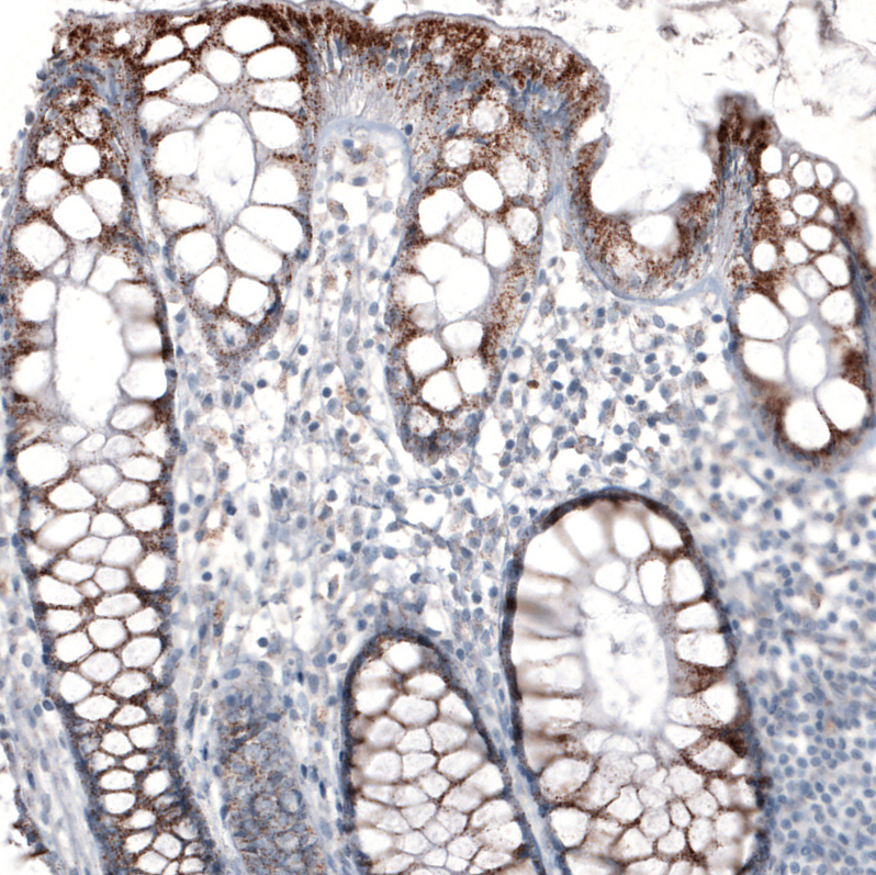
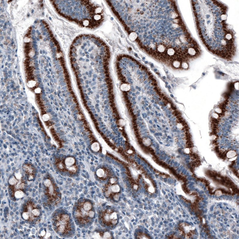
Immunohistochemical staining of human small intestine shows strong granular cytoplasmic positivity in glandular cells. |
|
|
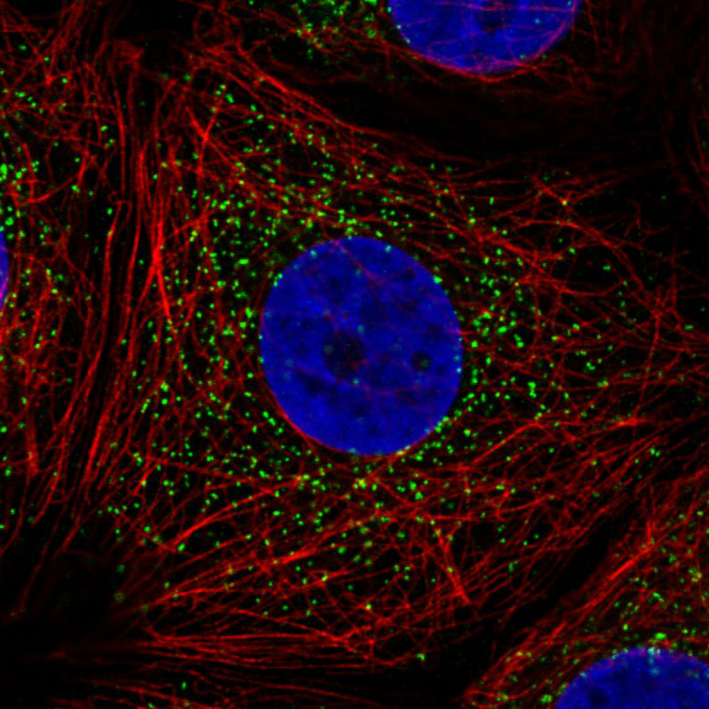
Immunofluorescence staining in MCF7 cell line with Anti-ABCD3 monoclonal antibody, showing specific staining of peroxisomes in green. Microtubule- and nuclear probes are visualized in red and blue respectively (where available). |
|
|
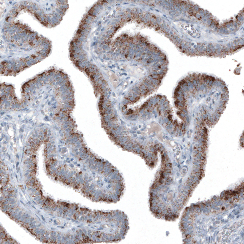
Immunohistochemical staining of human fallopian tube shows moderate granular cytoplasmic positivity in glandular cells. |
|
|
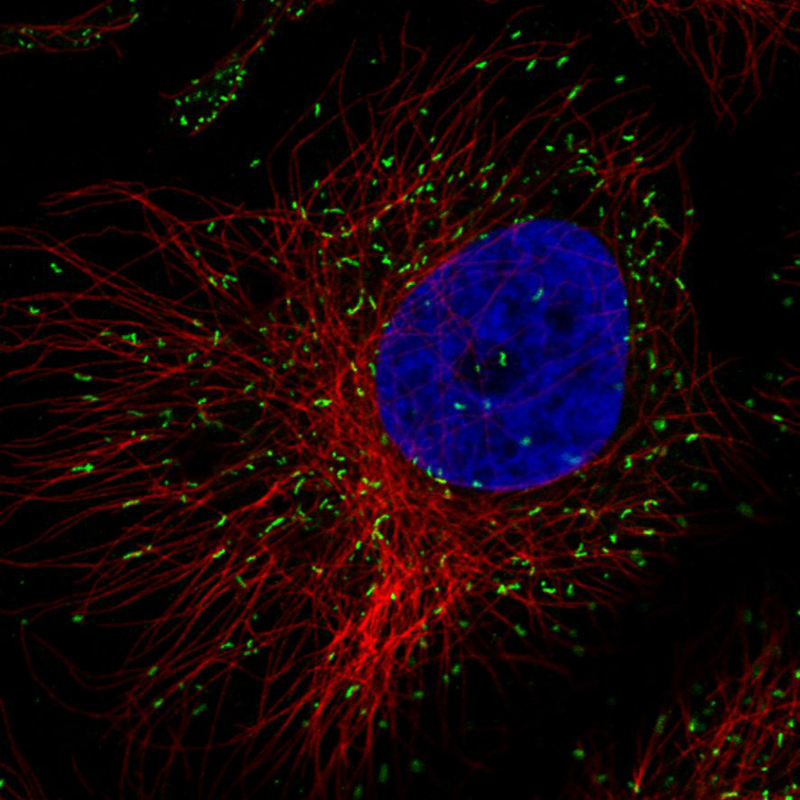
Immunofluorescence staining in HeLa cell line with Anti-ABCD3 monoclonal antibody, showing specific staining of peroxisomes in green. Microtubule- and nuclear probes are visualized in red and blue respectively (where available). |
|
|
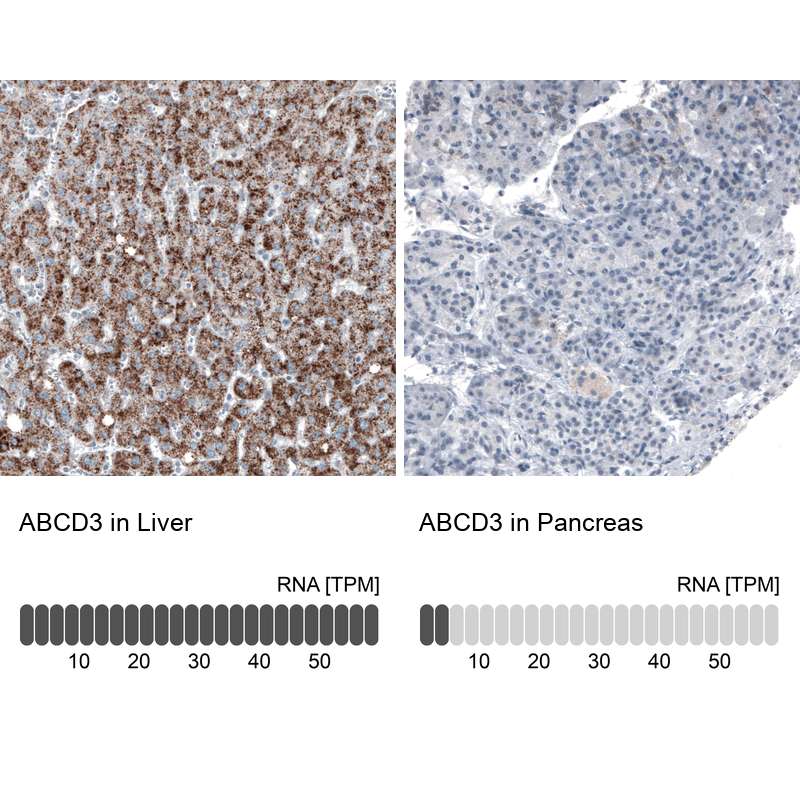
Immunohistochemistry analysis in human liver and pancreas tissues using AMAb90995 antibody. Corresponding ABCD3 RNA-seq data are presented for the same tissues. |
|
|
Immunofluorescence staining in A431 cell line with Anti-ABCD3 monoclonal antibody, showing specific staining of peroxisomes in green. Microtubule- and nuclear probes are visualized in red and blue respectively (where available). |
|
|
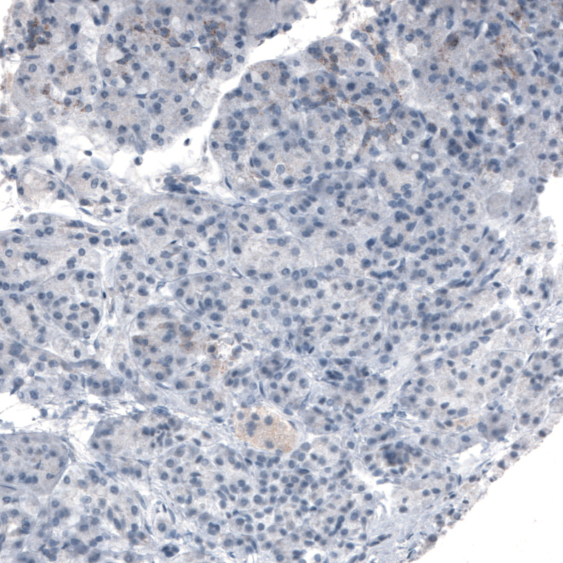
Immunohistochemical staining of human pancreas shows weak granular cytoplasmic positivity in exocrine glandular cells. |
|
|
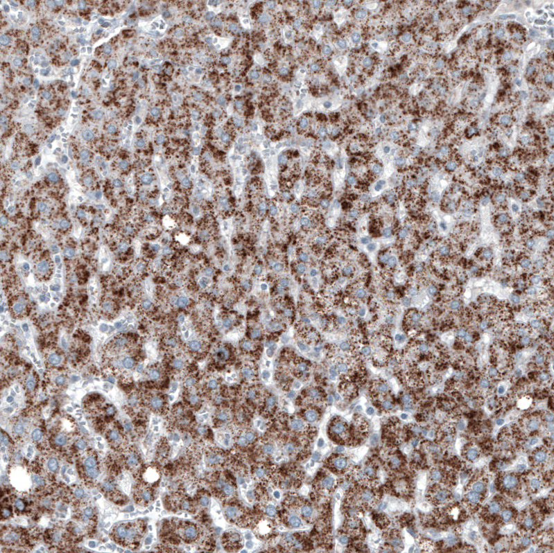
Immunohistochemical staining of human liver shows moderate granular cytoplasmic positivity in hepatocytes. |
Produktgarantie und fachkundiger Support
