
Anti-SORT1 Recombinant Antibody, IgG2b, Clone: [CL6526], Mouse, Monoclonal
Artikelnummer:
ATA-AMAB91427R
- Bilder (10)
| Artikelname: | Anti-SORT1 Recombinant Antibody, IgG2b, Clone: [CL6526], Mouse, Monoclonal |
| Artikelnummer: | ATA-AMAB91427R |
| Hersteller Artikelnummer: | AMAb91427R |
| Alternativnummer: | ATA-AMAB91427R-100, ATA-AMAB91427R-25 |
| Hersteller: | Atlas Antibodies |
| Wirt: | Mouse |
| Kategorie: | Antikörper |
| Applikation: | ICC, IHC, WB |
| Spezies Reaktivität: | Human, Mouse |
| Alternative Synonym: | Gp95, NT3 |
| Recombinant Mouse Monoclonal Anti-SORT1 Antibody against Human sortilin 1. Validated for Immunofluorescence, Immunohistochemistry and Western Blot |
|
|
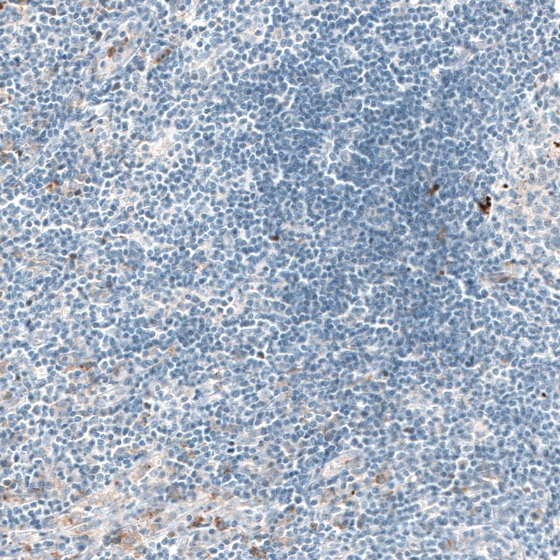
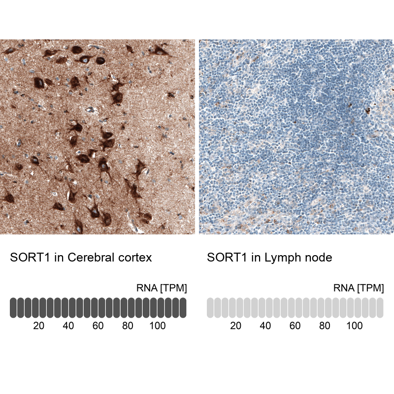
Immunohistochemical staining of human lymph node shows only weak positivity in a subset of lymphoid cells, as expected. |
|
|
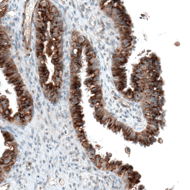
Immunohistochemical staining of human fallopian tube shows moderate to strong cytoplasmic positivity in glandular cells. |
|
|
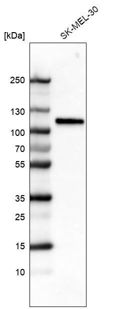
Western blot analysis in human cell line SK-MEL-30. |
|
|
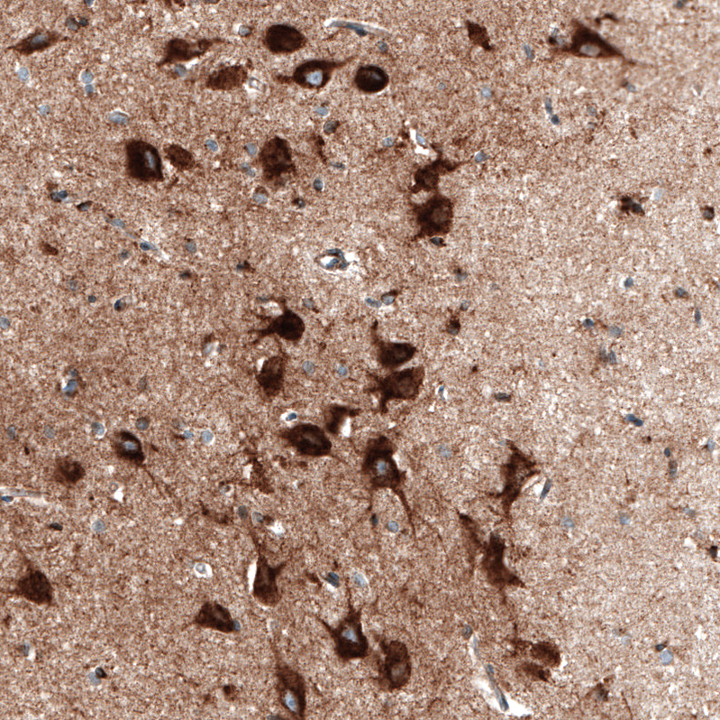
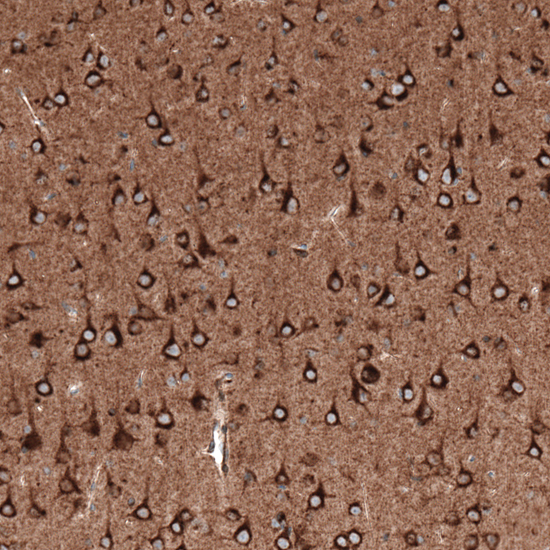
Immunohistochemical staining of human cerebral cortex shows strong cytoplasmic positivity in neurons. |
|
|
Immunohistochemical staining of mouse cerebral cortex shows strong cytoplasmic positivity in neurons. |
|
|
Immunohistochemistry analysis in human cerebral cortex and lymph node tissues using AMAb91427 antibody. Corresponding SORT1 RNA-seq data are presented for the same tissues. |
|
|
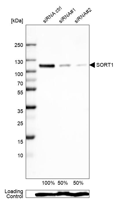
Western blot analysis in Rh30 cells transfected with control siRNA, target specific siRNA probe 1 and 2, using Anti-SORT1 antibody. Remaining relative intensity is presented. Loading control: Anti-GAPDH. |
|
|
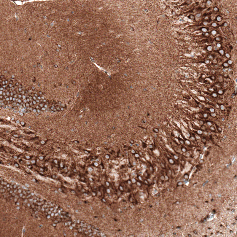
Immunohistochemical staining of mouse hippocampus shows strong cytoplasmic positivity in both pyramidal neurons and granular cells. |
|
|
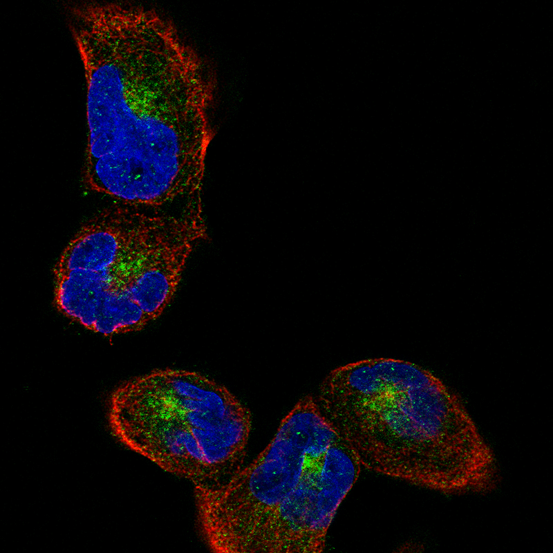
Immunofluorescence staining of RH-30 cells using the Anti-SORT1 monoclonal antibody, showing specific staining of the golgi apparatus in green. Microtubule- and nuclear probes are visualized in red and blue, respectively (where available). |
|
|
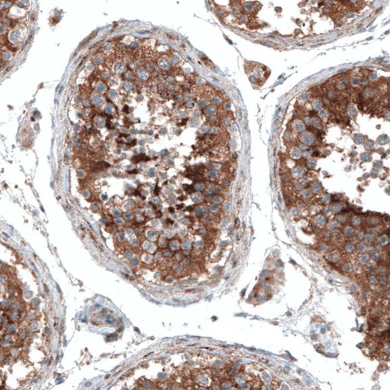
Immunohistochemical staining of human testis shows strong cytoplasmic positivity in cells in seminiferous ducts. |
Produktgarantie und fachkundiger Support
