Western blot, Optimal dilutions should be determined by end users. Immunohistochemistry (Paraffin-embedded Section), Optimal dilutions should be determined by end users. ELISA, Optimal dilutions should be determined by end users.
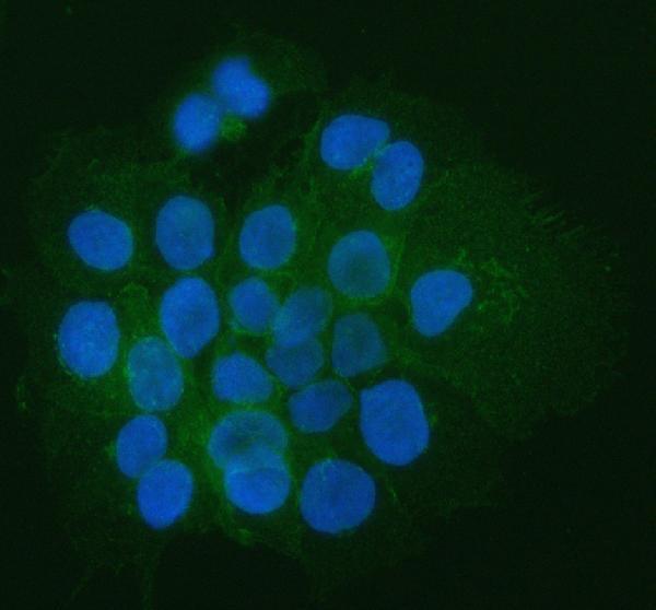
IF analysis of CD44 using anti-CD44 antibody (A00052-2). CD44 was detected in an immunocytochemical section of A431 cells. Enzyme antigen retrieval was performed using IHC enzyme antigen retrieval reagent (AR0022) for 15 mins. The cells were blocked with 10% goat serum. And then incubated with 5 µg/mL rabbit anti-CD44 Antibody (A00052-2) overnight at 4
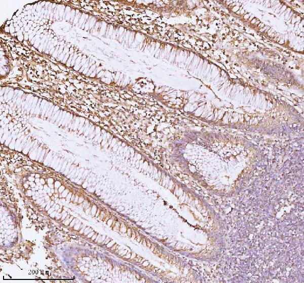
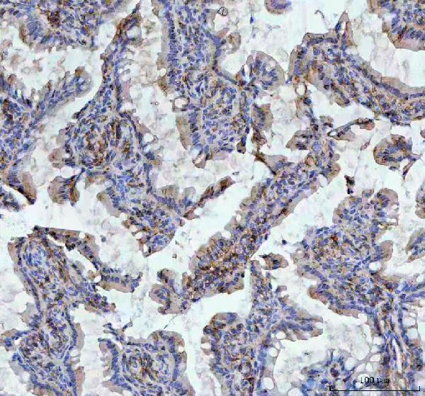
IHC analysis of CD44 using anti-CD44 antibody (A00052-2). CD44 was detected in a paraffin-embedded section of human colon tissue. Heat mediated antigen retrieval was performed in EDTA buffer (pH 8.0, epitope retrieval solution). The tissue section was blocked with 10% goat serum. The tissue section was then incubated with 2 µg/ml rabbit anti-CD44 Antibody (A00052-2) overnight at 4C. Peroxidase Conjugated Goat Anti-rabbit IgG was used as secondary antibody and incubated for 30 minutes at 37C. The tissue section was developed using HRP Conjugated Rabbit IgG Super Vision Assay Kit (Catalog SV0002) with DAB as the chromogen.
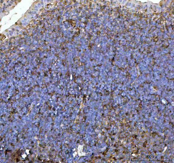
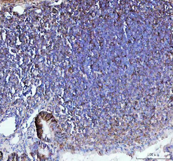
IHC analysis of CD44 using anti-CD44 antibody (A00052-2). CD44 was detected in a paraffin-embedded section of mouse lymphaden tissue. Heat mediated antigen retrieval was performed in EDTA buffer (pH 8.0, epitope retrieval solution). The tissue section was blocked with 10% goat serum. The tissue section was then incubated with 2 µg/ml rabbit anti-CD44 Antibody (A00052-2) overnight at 4C. Peroxidase Conjugated Goat Anti-rabbit IgG was used as secondary antibody and incubated for 30 minutes at 37C. The tissue section was developed using HRP Conjugated Rabbit IgG Super Vision Assay Kit (Catalog SV0002) with DAB as the chromogen.
IHC analysis of CD44 using anti-CD44 antibody (A00052-2). CD44 was detected in a paraffin-embedded section of rat colon tissue. Heat mediated antigen retrieval was performed in EDTA buffer (pH 8.0, epitope retrieval solution). The tissue section was blocked with 10% goat serum. The tissue section was then incubated with 2 µg/ml rabbit anti-CD44 Antibody (A00052-2) overnight at 4C. Peroxidase Conjugated Goat Anti-rabbit IgG was used as secondary antibody and incubated for 30 minutes at 37C. The tissue section was developed using HRP Conjugated Rabbit IgG Super Vision Assay Kit (Catalog SV0002) with DAB as the chromogen.
IHC analysis of CD44 using anti-CD44 antibody (A00052-2). CD44 was detected in a paraffin-embedded section of rat colon tissue. Heat mediated antigen retrieval was performed in EDTA buffer (pH 8.0, epitope retrieval solution). The tissue section was blocked with 10% goat serum. The tissue section was then incubated with 2 µg/ml rabbit anti-CD44 Antibody (A00052-2) overnight at 4C. Peroxidase Conjugated Goat Anti-rabbit IgG was used as secondary antibody and incubated for 30 minutes at 37C. The tissue section was developed using HRP Conjugated Rabbit IgG Super Vision Assay Kit (Catalog SV0002) with DAB as the chromogen.
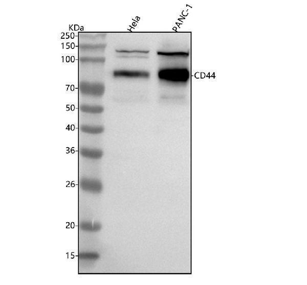
Western blot analysis of CD44 using anti-CD44 antibody (A00052-2). Electrophoresis was performed on a 5-20% SDS-PAGE gel at 70V (Stacking gel) / 90V (Resolving gel) for 2-3 hours. The sample well of each lane was loaded with 30 ug of sample under reducing conditions. Lane 1: human Hela whole cell lysates,Lane 2: human PANC-1 whole cell lysates.After electrophoresis, proteins were transferred to a nitrocellulose membrane at 150 mA for 50-90 minutes. Blocked the membrane with 5% non-fat milk/TBS for 1.5 hour at RT. The membrane was incubated with rabbit anti-CD44 antigen affinity purified polyclonal antibody (Catalog A00052-2) at 0.5 µg/mL overnight at 4C, then washed with TBS-0.1%Tween 3 times with 5 minutes each and probed with a goat anti-rabbit IgG-HRP secondary antibody at a dilution of 1:5000 for 1.5 hour at RT. The signal is developed using an Enhanced Chemiluminescent detection (ECL) kit (Catalog EK1002) with Tanon 5200 system. A specific band was detected for CD44 at approximately 85 kDa. The expected band size for CD44 is at 82 kDa.
* Mehrwertsteuer und Versandkosten nicht enthalten. Irrtümer und Preisänderungen vorbehalten