500 µg/ml antibody with PBS, 0.02% NaN3, 1 mg stabilizing protein and 50% glycerolThis antibody is supplied in a stabilized formulation. Compatibility with conjugation reactions depends on the chemistry of the conjugation method used. For conjugation meth
Formulierung:
Liquid
Application Verdünnung:
Western blot, 1:500-2000Immunohistochemistry, 1:50-400Immunocytochemistry/Immunofluorescence, 1:50-400Immunoprecipitation, 1:250-300ELISA, 1:100-1000
Upregulation of PITX1 in hypoxic mice and PASMCs. A , B Immunofluorescence staining: Localization and statistical data of PITX1 in tissues. Scale bar: 25µm. C Western blot: Representative image and statistical data of PITX1 expression in the lung tissues of mice. D RT‒qPCR: Changes in the transcript level of PITX1 in the lung tissues of mice. E Western blot: Representative image and statistical data of PITX1 expression in the smooth muscle cells of mice. F RT‒qPCR: Changes in the transcript level of PITX1 in the smooth muscle cells of mice. Nor normoxic, Hyp hypoxic. All values are presented as meansSEMs (* p <0.05, ** p <0.01, and *** p <0.001, n3) Index in PubMed under a CC BY license. PMID: 40241046
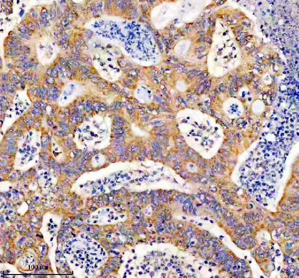
IHC analysis of PITX1 using anti-PITX1 antibody (A02993-2). PITX1 was detected in a paraffin-embedded section of human colon cancer tissue. Heat mediated antigen retrieval was performed in EDTA buffer (pH 8.0, epitope retrieval solution). The tissue section was blocked with 10% goat serum. The tissue section was then incubated with 1:200 rabbit anti-PITX1 Antibody (A02993-2) overnight at 4C. Peroxidase Conjugated Goat Anti-rabbit IgG was used as secondary antibody and incubated for 30 minutes at 37C. The tissue section was developed using HRP Conjugated Rabbit IgG Super Vision Assay Kit (Catalog SV0002) with DAB as the chromogen.
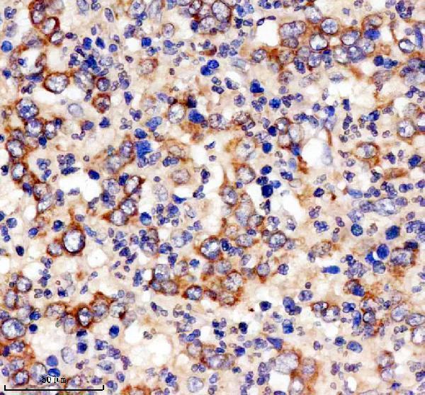
IHC analysis of PITX1 using anti-PITX1 antibody (A02993-2). PITX1 was detected in a paraffin-embedded section of human stomach cancer tissue. Heat mediated antigen retrieval was performed in EDTA buffer (pH 8.0, epitope retrieval solution). The tissue section was blocked with 10% goat serum. The tissue section was then incubated with 1:200 rabbit anti-PITX1 Antibody (A02993-2) overnight at 4C. Peroxidase Conjugated Goat Anti-rabbit IgG was used as secondary antibody and incubated for 30 minutes at 37C. The tissue section was developed using HRP Conjugated Rabbit IgG Super Vision Assay Kit (Catalog SV0002) with DAB as the chromogen.
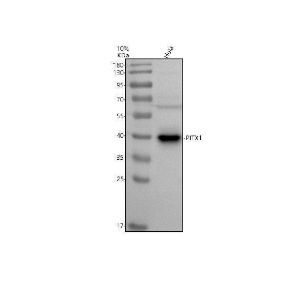
Western blot analysis of PITX1 using anti-PITX1 antibody (A02993-2). Electrophoresis was performed on a 10% SDS-PAGE gel at 80V (Stacking gel) / 120V (Resolving gel) for 2 hours. The sample well of each lane was loaded with 30 ug of sample under reducing conditions. Lane 1: human Hela whole cell lysates.After electrophoresis, proteins were transferred to a nitrocellulose membrane at 150 mA for 50-90 minutes. Blocked the membrane with 5% non-fat milk/TBS for 1.5 hour at RT. The membrane was incubated with rabbit anti-PITX1 antigen affinity purified polyclonal antibody (A02993-2) at 1:1000 overnight at 4C, then washed with TBS-0.1%Tween 3 times with 5 minutes each and probed with a goat anti-rabbit IgG-HRP secondary antibody at a dilution of 1:5000 for 1.5 hour at RT. The signal is developed using an ECL Plus Western Blotting Substrate (Catalog AR1196-200) with Tanon 5200 system. A specific band was detected for PITX1 at approximately 40 kDa. The expected band size for PITX1 is at 34 kDa.
Impact of PITX1 on pyroptosis in hypoxia-induced PASMCs. A GSEA: The functions of PITX1 were enriched mainly in the inflammasome signaling pathway under hypoxic conditions. B , C Western blot: The interference efficiency of siPITX1 and the overexpression efficiency of the PITX1 plasmid. D , I Western blot: Representative images and statistical data of CASP1, GSDMD-N, IL-1beta, and IL-18 levels. E , J YPI/PI staining: YPI is a green fluorescent dye permeable to the membrane of apoptotic cells, and PI staining of necrotic cells with compromised membrane integrity may result in the emission of red fluorescence. Scale bar: 100µm. F , K DiO staining: Green fluorescence staining of the cell membrane, and the membranes of live cells exhibit green fluorescence. Scale bar: 50µm. G , H LDH assay: Determination of the number of dead cells by measuring the amou
* Mehrwertsteuer und Versandkosten nicht enthalten. Irrtümer und Preisänderungen vorbehalten