Boster Bio Anti-IRF3 Rabbit Monoclonal Antibody catalog M00165. Tested in WB, IHC, ICC/IF, Flow Cytometry applications. This antibody reacts with Human, Mouse, Rat.
Rabbit IgG in stabilizing components, phosphate buffered saline, pH 7.4, 150mM NaCl, 0.02% sodium azide and 50% glycerol. *This antibody is supplied in a stabilized formulation. Compatibility with conjugation reactions depends on the chemistry of the con
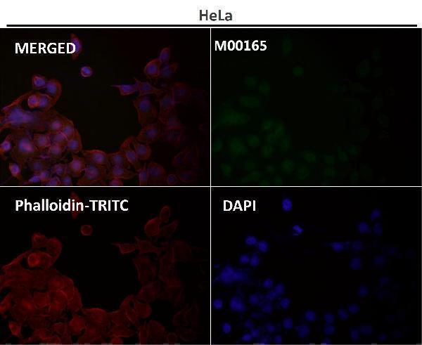
Immunofluorescent analysis using the Antibody at 1:50 dilution.
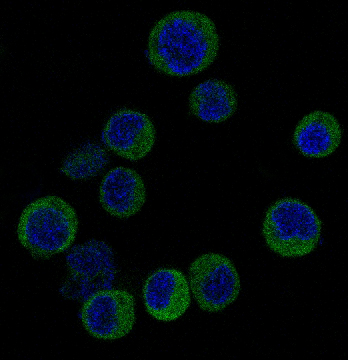
Immunofluorescent analysis of Jurkat cells, using IRF3 Antibody.
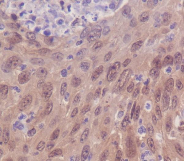
Immunohistochemical analysis of paraffin-embedded human cervix carcinoma, using IRF3 Antibody.
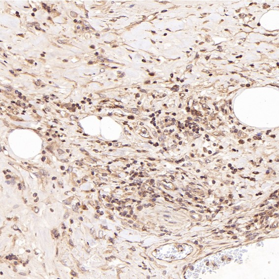
Immunohistochemical analysis of paraffin-embedded Human Hodgkins lymphoma, using the Antibody at 1:50 dilution.
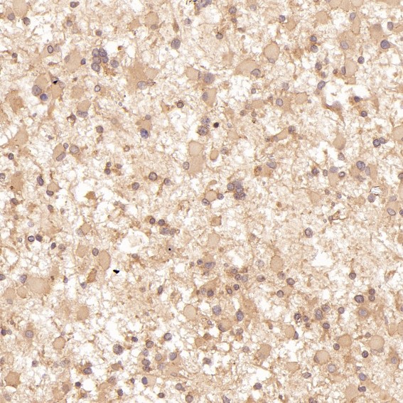
Immunohistochemical analysis of paraffin-embedded Human astrocytoma, using the Antibody at 1:50 dilution.
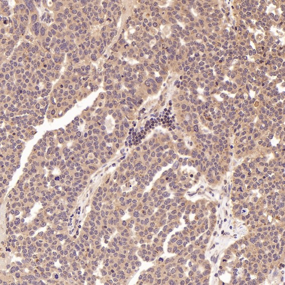
Immunohistochemical analysis of paraffin-embedded Human ovarian cancer, using the Antibody at 1:50 dilution.
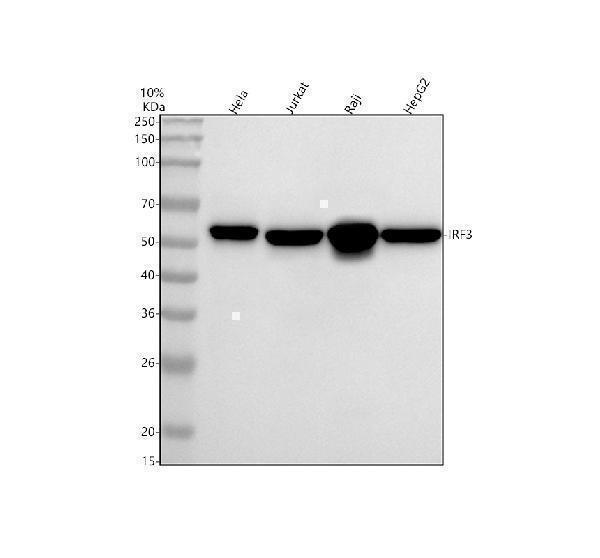
Western blot analysis of IRF3 using anti-IRF3 antibody (M00165). Electrophoresis was performed on a 10% SDS-PAGE gel at 80V (Stacking gel) / 120V (Resolving gel) for 2 hours. The sample well of each lane was loaded with 30 ug of sample under reducing conditions. Lane 1: human Hela whole cell lysates,Lane 2: human Jurkat whole cell lysates,Lane 3: human Raji whole cell lysates,Lane 4: human HepG2 whole cell lysates.After electrophoresis, proteins were transferred to a nitrocellulose membrane at 150 mA for 50-90 minutes. Blocked the membrane with 5% non-fat milk/TBS for 1.5 hour at RT. The membrane was incubated with rabbit anti-IRF3 antigen affinity purified monoclonal antibody (M00165) at 1:500 overnight at 4C, then washed with TBS-0.1%Tween 3 times with 5 minutes each and probed with a goat anti-rabbit IgG-HRP secondary antibody at a dilution of 1:5000 for 1.5 hour at RT. The signal is developed using an ECL Plus Western Blotting Substrate (Catalog AR1196-200) with Tanon 5200 system. A specific band was detected for IRF3 at approximately 60 kDa. The expected band size for IRF3 is at 47 kDa.
All lanes use the Antibody at 1:2W dilution for 1 hour at room temperature.
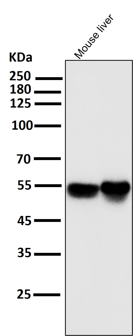
Western blot analysis of IRF3 using anti-IRF3 antibody (M00165). Electrophoresis was performed on a 10% SDS-PAGE gel at 80V (Stacking gel) / 120V (Resolving gel) for 2 hours. The sample well of each lane was loaded with 30 ug of sample under reducing conditions. Lane 1: human A549- WT whole cell lysates,Lane 2: human A549-IRF3 KO whole cell lysates.After electrophoresis, proteins were transferred to a nitrocellulose membrane at 150 mA for 50-90 minutes. Blocked the membrane with 5% non-fat milk/TBS for 1.5 hour at RT. The membrane was incubated with rabbit anti-IRF3 antigen affinity purified monoclonal antibody (M00165) at 0.5 µg/mL overnight at 4C, then washed with TBS-0.1%Tween 3 times with 5 minutes each and probed with a goat anti-rabbit IgG-HRP secondary antibody at a dilution of 1:5000 for 1.5 hour at RT. The signal is developed using an ECL Plus Western Blotting Substrate (Catalog AR1196-200) with Tanon 5200 system. A specific band was detected for IRF3 at approximately 60 kDa. The expected band size for IRF3 is at 47 kDa.
* Mehrwertsteuer und Versandkosten nicht enthalten. Irrtümer und Preisänderungen vorbehalten