Boster Bio Anti-TMPRSS2 Rabbit Monoclonal Antibody catalog M00666. Tested in WB, IHC, ICC/IF, Flow Cytometry applications. This antibody reacts with Human, Mouse, Rat.
Rabbit IgG in stabilizing components, phosphate buffered saline, pH 7.4, 150mM NaCl, 0.02% sodium azide and 50% glycerol. *This antibody is supplied in a stabilized formulation. Compatibility with conjugation reactions depends on the chemistry of the con
Reinheit:
Affinity-chromatography
Formulierung:
Liquid
Target-Kategorie:
Transmembrane protease serine 2
Application Verdünnung:
WB 1:500-2000IHC 1:50-200ICC/IF 1:50-200FC 1:60
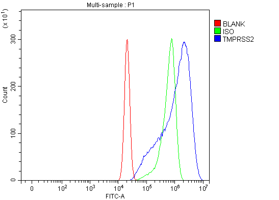
Flow Cytometry analysis of U2OS cells using anti-TMPRSS2 antibody (M00666). Overlay histogram showing U2OS cells stained with M00666 (Blue line). The cells were blocked with 10% normal goat serum. And then incubated with mouse anti-TMPRSS2 Antibody (M00666) at 1:60 dilution for 30 min at 20C. DyLight488 conjugated goat anti-mouse IgG (BA1126) at 1:60 was used as secondary antibody for 30 minutes at 20C. Isotype control antibody (Green line) was mouse IgG at 1:60 used under the same conditions. Unlabelled sample (Red line) was also used as a control.
Western blot analysis of TMPRSS2 using anti-TMPRSS2 antibody (M00666). Electrophoresis was performed on a 5-20% SDS-PAGE gel at 70V (Stacking gel) / 90V (Resolving gel) for 2-3 hours. The sample well of each lane was loaded with 30 ug of sample under reducing conditions. Lane 1: human LNCAP whole cell lysates,Lane 2: human COLO-320 whole cell lysates,Lane 3: human RT4 whole cell lysates,Lane 4: human Caco-2 whole cell lysates,Lane 5: rat kidney tissue lysates.After electrophoresis, proteins were transferred to a nitrocellulose membrane at 150 mA for 50-90 minutes. Blocked the membrane with 5% non-fat milk/TBS for 1.5 hour at RT. The membrane was incubated with rabbit anti-TMPRSS2 antigen affinity purified monoclonal antibody (Catalog M00666) at 1:500 overnight at 4C, then washed with TBS-0.1%Tween 3 times with 5 minutes each and probed with a goat anti-rabbit IgG-HRP secondary antibody at a dilution of 1:5000 for 1.5 hour at RT. The signal is developed using an Enhanced Chemiluminescent detection (ECL) kit (Catalog EK1002) with Tanon 5200 system. A specific band was detected for TMPRSS2 at approximately 54 kDa. The expected band size for TMPRSS2 is at 54 kDa.
All lanes use the Antibody at 1:1K dilution for 1 hour at room temperature.
All lanes use the Antibody at 1:1K dilution for 1 hour at room temperature.
* Mehrwertsteuer und Versandkosten nicht enthalten. Irrtümer und Preisänderungen vorbehalten