
Anti-CD79B Mouse Monoclonal Antibody [Clone ID: OTI8A12], IgG1, Clone: [Clone: OTI8A12]
Artikelnummer:
BOB-M01399-2
- Bilder (9)


| Artikelname: | Anti-CD79B Mouse Monoclonal Antibody [Clone ID: OTI8A12], IgG1, Clone: [Clone: OTI8A12] |
| Artikelnummer: | BOB-M01399-2 |
| Hersteller Artikelnummer: | M01399-2 |
| Alternativnummer: | BOB-M01399-2-100UL |
| Hersteller: | Boster Bio |
| Wirt: | Mouse |
| Kategorie: | Antikörper |
| Applikation: | FC, IHC, WB |
| Spezies Reaktivität: | Human |
| Immunogen: | Full length human recombinant protein of human CD79B (NP_001035022) produced in HEK293T. |
| Boster Bio CD79B mouse monoclonal antibody,clone OTI8A12. Catalog M01399-2. Tested in FC, IHC, WB. This antibody reacts with Human. |
| Klonalität: | Monoclonal |
| Konzentration: | 1 mg/ml |
| Klon-Bezeichnung: | [Clone: OTI8A12] |
| Isotyp: | IgG1 |
| UniProt: | P40259 |
| Puffer: | PBS (pH 7.3) containing 1% stabilizing protein, 50% glycerol and 0.02% sodium azide.This antibody is supplied in a stabilized formulation. Compatibility with conjugation reactions depends on the chemistry of the conjugation method used. For conjugation me |
| Application Verdünnung: | WB 1:500~2000IHC 1:2000 |
|
|
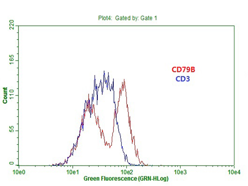
Flow cytometric Analysis of Ramos cells |
|
|
Flow cytometric Analysis of Jurkat cells |
|
|
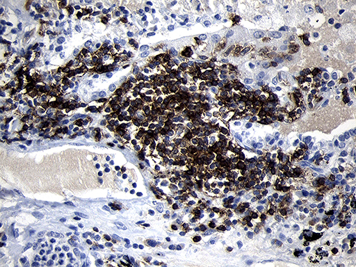
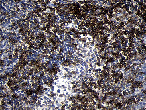
Immunohistochemical staining of paraffin-embedded Carcinoma of Human lung tissue using anti-CD79B mouse monoclonal antibody. (Heat-induced epitope retrieval by 1mM EDTA in 10mM Tris buffer (pH8.5) at 120AC for 3min |
|
|
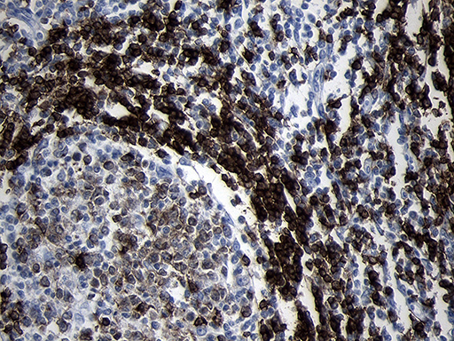
Immunohistochemical staining of paraffin-embedded Human lymph node tissue within the normal limits using anti-CD79B mouse monoclonal antibody. (Heat-induced epitope retrieval by 1mM EDTA in 10mM Tris buffer (pH8.5) at 120AC for 3min |
|
|
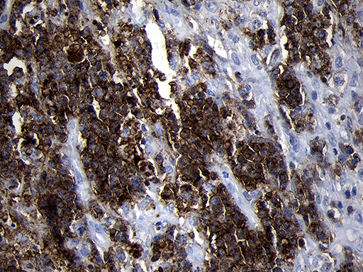
Immunohistochemical staining of paraffin-embedded Human lymphoma tissue using anti-CD79B mouse monoclonal antibody. (Heat-induced epitope retrieval by 1mM EDTA in 10mM Tris buffer (pH8.5) at 120AC for 3min |
|
|
Immunohistochemical staining of paraffin-embedded Human tonsil within the normal limits using anti-CD79B mouse monoclonal antibody. (Heat-induced epitope retrieval by 1mM EDTA in 10mM Tris buffer (pH8.5) at 120AC for 3min |
|
|
HEK293T cells were transfected with the pCMV6-ENTRY control (Left lane) or pCMV6-ENTRY CD79B (Right lane) cDNA for 48 hrs and lysed. Equivalent amounts of cell lysates (5 ug per lane) were separated by SDS-PAGE and immunoblotted with anti-CD79B (1:2000). |
|
|
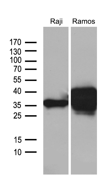
Western blot analysis of extracts (35ug) from 2 different cell lines by using anti-CD79B monoclonal antibody (1:500). |
|
|
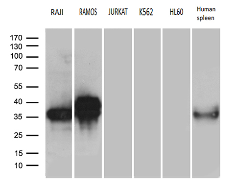
Western blot analysis of extracts (35ug) from 5 different cell lines and human spleen tissue lysate by using anti-CD79A monoclonal antibody (1:500). |
Produktgarantie und fachkundiger Support
