
Cyclin B1 Rabbit Polyclonal Antibody, Unconjugated
Artikelnummer:
BYT-ORB10494
- Bilder (9)


| Artikelname: | Cyclin B1 Rabbit Polyclonal Antibody, Unconjugated |
| Artikelnummer: | BYT-ORB10494 |
| Hersteller Artikelnummer: | orb10494 |
| Alternativnummer: | BYT-ORB10494-100,BYT-ORB10494-200,BYT-ORB10494-50 |
| Hersteller: | Biorbyt |
| Wirt: | Rabbit |
| Kategorie: | Antikörper |
| Applikation: | FC, ICC, IF, IHC-Fr, IHC-P, WB |
| Spezies Reaktivität: | Human, Mouse, Rat |
| Immunogen: | KLH conjugated synthetic peptide derived from human Cyclin B1 (271-433/433aa) |
| Konjugation: | Unconjugated |
| Alternative Synonym: | CCNB 1, CCNB, CCNB1, CCNB1_HUMAN, G2 mitotic specific cyclin B1, G2/mitotic-specific cyclin-B1. |
| Cyclin B1 Rabbit Polyclonal Antibody |
| Application Verdünnung: | WB=1:500-2000, IHC-P=1:100-500, IHC-F=1:100-500, ICC/IF=1:100-500, IF=1:100-500, Flow-Cyt=1µg/Test |
|
|
Paraformaldehyde-fixed, paraffin embedded (Mouse small intestine), Antigen retrieval by boiling in sodium citrate buffer (pH6.0) for 15 min, Block endogenous peroxidase by 3% hydrogen peroxide for 20 minutes, Blocking buffer (normal goat serum) at 37C for 30 min, Antibody incubation with (Cyclin B1) Polyclonal Antibody, Unconjugated (orb10494) at 1:400 overnight at 4C, followed by operating according to SP Kit (Rabbit) instructionsand DAB staining. |
|
|
Paraformaldehyde-fixed, paraffin embedded (Rat esophageal), Antigen retrieval by boiling in sodium citrate buffer (pH6.0) for 15 min, Block endogenous peroxidase by 3% hydrogen peroxide for 20 minutes, Blocking buffer (normal goat serum) at 37C for 30 min, Antibody incubation with (Cyclin B1) Polyclonal Antibody, Unconjugated (orb10494) at 1:400 overnight at 4C, followed by operating according to SP Kit (Rabbit) instructionsand DAB staining. |
|
|
Sample: Lane 1: Lymph node (Rat) Lysate at 40 ug, Lane 2: Testis (Rat) Lysate at 40 ug, Lane 3: Spleen (Rat) Lysate at 40 ug, Primary: Anti-Cyclin B1 (orb10494) at 1/1000 dilution, Secondary: IRDye800CW Goat Anti-Rabbit IgG at 1/20000 dilution, Predicted band size: 55-60 kD, Observed band size: 60 kD. |
|
|
Sample: U251 Cell (Human) Lysate at 30 ug, Primary: Anti-Cyclin B1 (orb10494) at 1/300 dilution, Secondary: IRDye800CW Goat Anti-Rabbit IgG at 1/20000 dilution, Predicted band size: 48 kD, Observed band size: 50 kD. |
|
|
Sample: U937 Cell (Human) Lysate at 30 ug, Primary: Anti-Cyclin B1 (orb10494) at 1/300 dilution, Secondary: IRDye800CW Goat Anti-Rabbit IgG at 1/20000 dilution, Predicted band size: 48 kD, Observed band size: 50 kD. |
|
|
Tissue/Cell: human colon carcinoma, 4% Paraformaldehyde-fixed and paraffin-embedded, Antigen retrieval: citrate buffer (0.01M, pH6.0), Boiling bathing for 15 min, Block endogenous peroxidase by 3% Hydrogen peroxide for 30 min, Blocking buffer (normal goat serum) at 37C for 20 min, Incubation: Anti-Cyclin B1 Polyclonal Antibody, Unconjugated (orb10494) 1:200, overnight at 4C, followed by conjugation to the secondary antibody and DAB staining. |
|
|
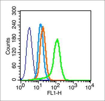
Blank control (blue line): A549 (blue). Primary Antibody (green line): Rabbit Anti-Cyclin B1 antibody (orb10494). Dilution: 1 µg/10 6 cells, Isotype Control Antibody (orange line): Rabbit IgG. Secondary Antibody (white blue line): F (ab)2 fragment goat anti-rabbit IgG-FITC. Dilution: 1 µg/Test. Protocol, The cells were fixed with 2% paraformaldehyde (10 min) and then permeabilized with 0.1% PBS-Tween for 20 min at room temperature. Cells stained with Primary Antibody for 30 min at room temperature. The cells were then incubated in 1 X PBS/2% BSA/10% goat serum to block non-specific protein-protein interactions followed by the antibody for 15 min at room temperature. The secondary antibody used for 40 min at room temperature. Acquisition of 20000 events was performed. |
|
|
Cell: Hela, Concentration: 1:100, Host/Isotype: Rabbit/IgG, Flow cytometric analysis of primary antibody (Cat: orb10494) on Hela (green) compared with isotype control in the absence of primary antibody (blue) followed by Alexa Fluor 488-conjugated goat anti-rabbit IgG (H+L) secondary antibody. |
|
|
Hela cell, 4% Paraformaldehyde-fixed, Triton X-100 at room temperature for 20 min, Blocking buffer (normal goat serum) at 37C for 20 min, Antibody incubation with (Cyclin B1) polyclonal Antibody, Unconjugated (orb10494) 1:100, 90 minutes at 37C, followed by a conjugated Goat Anti-Rabbit IgG antibody at 37C for 90 minutes, DAPI (blue) was used to stain the cell nuclei. |
Produktgarantie und fachkundiger Support
