Application Notes: 1 µg/ml was sufficient for detection of TNFR1 in 20 µg of Hela lysate by colorimetric immunoblot analysis using Goat anti-rabbit IgG:HRP as the secondary antibody
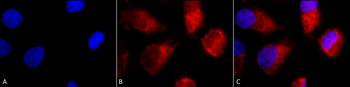
Immunocytochemistry/Immunofluorescence analysis using Rabbit Anti-TNF-R1 Polyclonal Antibody. Tissue: Cervical cancer cell line (HeLa). Species: Human. Fixation: 2% Formaldehyde for 20 min at RT. Primary Antibody: Rabbit Anti-TNF-R1 Polyclonal Antibody at 1:100 for 12 hours at 4C. Secondary Antibody: APC Goat Anti-Rabbit (red) at 1:200 for 2 hours at RT. Counterstain: DAPI (blue) nuclear stain at 1:40000 for 2 hours at RT. Localization: Golgi apparatus membrane. Magnification: 100x. (A) DAPI (blue) nuclear stain. (B) Anti-TNF-R1 Antibody. (C) Composite.
Immunohistochemistry analysis using Rabbit Anti-TNF-R1 Polyclonal Antibody. Tissue: backskin. Species: Mouse. Fixation: Bouins Fixative Solution. Primary Antibody: Rabbit Anti-TNF-R1 Polyclonal Antibody at 1:100 for 1 hour at RT. Secondary Antibody: FITC Goat Anti-Rabbit (green) at 1:50 for 1 hour at RT. Localization: dermis.
Western blot analysis of Mouse Liver cell lysates showing detection of ~55 kDa TNF-R1 protein using Rabbit Anti-TNF-R1 Polyclonal Antibody. Lane 1: Molecular Weight Ladder (MW). Lane 2: Mouse Liver cell lysates. Load: 15 µg. Block: 5% Skim Milk in 1X TBST. Primary Antibody: Rabbit Anti-TNF-R1 Polyclonal Antibody at 1:1000 for 2 hours at RT. Secondary Antibody: Goat Anti-Rabbit IgG: HRP at 1:2000 for 60 min at RT. Color Development: ECL solution for 5 min at RT. Predicted/Observed Size: ~55 kDa.
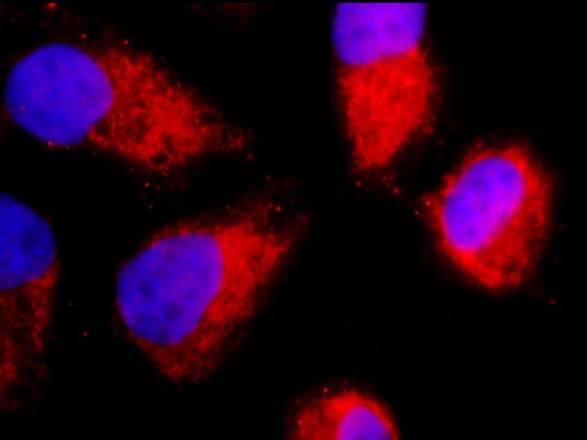
Immunocytochemistry/Immunofluorescence analysis using Rabbit Anti-TNF-R1 Polyclonal Antibody. Tissue: Cervical cancer cell line (HeLa). Species: Human. Fixation: 2% Formaldehyde for 20 min at RT. Primary Antibody: Rabbit Anti-TNF-R1 Polyclonal Antibody at 1:100 for 12 hours at 4C. Secondary Antibody: FITC Goat Anti-Rabbit (green) at 1:200 for 2 hours at RT. Counterstain: DAPI (blue) nuclear stain at 1:40000 for 2 hours at RT. Localization: Golgi apparatus membrane. Magnification: 20x. (A) DAPI (blue) nuclear stain. (B) Anti-TNF-R1 Antibody. (C) Composite.
Western blot analysis of Human A549 showing detection of ~ 50 kDa TNF-R1 protein using Rabbit Anti-TNF-R1 Polyclonal Antibody. Lane 1: MW Ladder, Lane 2: A549. Load: 30 ug. Block: 5% BSA in TBST. Primary Antibody: Rabbit Anti-TNF-R1 Polyclonal Antibody at 1:1000 for 2 hours at RT with shaking. Secondary Antibody: Goat Anti-Rabbit IgG: HRP at 1:4000 for 1 hour at RT with shaking. Color Development: Chemiluminescent for HRP (Moss) for 5 min in RT. Predicted/Observed Size: ~ 50 kDa. Other Band (s): ~90-100kDa. Other bands can be explained by a few factors, such as oligomerization, self-aggregation, cleavage of the TNFR1 extracellular domain, etc.
Immunocytochemistry/Immunofluorescence analysis using Rabbit Anti-TNF-R1 Polyclonal Antibody. Tissue: HaCaT cells. Species: Human. Fixation: Cold 100% methanol at -20C for 10 minutes. Primary Antibody: Rabbit Anti-TNF-R1 Polyclonal Antibody at 1:100 for 12 hours at 4C. Secondary Antibody: FITC Goat Anti-Rabbit at 1:50 for 1-2 hours at RT in dark. Localization: Punctate nuclear staining, dotty staining in cytoplasm.
Immunofluorescence analysis of hela cells using TNF-R1 antibody
Immunofluorescence analysis of hela cells using TNF-R1 antibody
* Mehrwertsteuer und Versandkosten nicht enthalten. Irrtümer und Preisänderungen vorbehalten