A synthetic peptide corresponding to a sequence in the middle region of human KRT7, which shares 91.3% amino acid (aa) sequence identity with mouse and rat KRT7.
Konjugation:
Unconjugated
Alternative Synonym:
Cell and organelle markers, CK 7, CK7, Cytokeratin 7, Cytokeratin 7-specific, Cytoskeleton Marker, K2C7, K7, keratin 7, KRT7, Sarcolectin, SCL, Type II keratin Kb7
Anti-KRT7 Antibody. Tested in IHC, WB applications. This antibody reacts with Human.
Klonalität:
Polyclonal
Konzentration:
Adding 0.2 ml of distilled water will yield a concentration of 500 µg/ml.
Western blot, 0.25-0.5 µg/ml, Human Immunohistochemistry(Paraffin-embedded Section), 2-5 µg/ml, Human
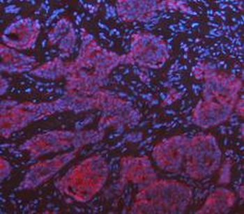
IF analysis of KRT7 using anti-KRT7 antibody. KRT7 was detected in a paraffin-embedded section of human breast cancer tissue. Heat mediated antigen retrieval was performed in EDTA buffer (pH8.0, epitope retrieval solution). The tissue section was blocked with 10% goat serum. The tissue section was then incubated with 25 µg/mL rabbit anti-KRT7 Antibody overnight at 4C. DyLight 594 Conjugated AffiniPure Goat Anti-rabbit IgG(H+L) was used as secondary antibody at 1:100 dilution and incubated for 30 minutes at 37C. The section was counterstained with DAPI. Visualize using a fluorescence microscope and filter sets appropriate for the label used.
IF analysis of KRT7 using anti-KRT7 antibody. KRT7 was detected in a paraffin-embedded section of human lung cancer tissue. Heat mediated antigen retrieval was performed in EDTA buffer (pH8.0, epitope retrieval solution). The tissue section was blocked with 10% goat serum. The tissue section was then incubated with 25 µg/mL rabbit anti-KRT7 Antibody overnight at 4C. DyLight 594 Conjugated AffiniPure Goat Anti-rabbit IgG(H+L) was used as secondary antibody at 1:100 dilution and incubated for 30 minutes at 37C. The section was counterstained with DAPI. Visualize using a fluorescence microscope and filter sets appropriate for the label used.
IF analysis of KRT7 using anti-KRT7 antibody. KRT7 was detected in a paraffin-embedded section of human ovarian cancer tissue. Heat mediated antigen retrieval was performed in EDTA buffer (pH8.0, epitope retrieval solution). The tissue section was blocked with 10% goat serum. The tissue section was then incubated with 25 µg/mL rabbit anti-KRT7 Antibody overnight at 4C. DyLight 594 Conjugated AffiniPure Goat Anti-rabbit IgG(H+L) was used as secondary antibody at 1:100 dilution and incubated for 30 minutes at 37C. The section was counterstained with DAPI. Visualize using a fluorescence microscope and filter sets appropriate for the label used.
IF analysis of KRT7 using anti-KRT7 antibody. KRT7 was detected in a paraffin-embedded section of human stomach cancer tissue. Heat mediated antigen retrieval was performed in EDTA buffer (pH8.0, epitope retrieval solution). The tissue section was blocked with 10% goat serum. The tissue section was then incubated with 25 µg/mL rabbit anti-KRT7 Antibody overnight at 4C. DyLight 594 Conjugated AffiniPure Goat Anti-rabbit IgG(H+L) was used as secondary antibody at 1:100 dilution and incubated for 30 minutes at 37C. The section was counterstained with DAPI. Visualize using a fluorescence microscope and filter sets appropriate for the label used.
IF analysis of KRT7 using anti-KRT7 antibody. KRT7 was detected in a paraffin-embedded section of human throid cancer tissue. Heat mediated antigen retrieval was performed in EDTA buffer (pH8.0, epitope retrieval solution). The tissue section was blocked with 10% goat serum. The tissue section was then incubated with 25 µg/mL rabbit anti-KRT7 Antibody overnight at 4C. DyLight 594 Conjugated AffiniPure Goat Anti-rabbit IgG(H+L) was used as secondary antibody at 1:100 dilution and incubated for 30 minutes at 37C. The section was counterstained with DAPI. Visualize using a fluorescence microscope and filter sets appropriate for the label used.
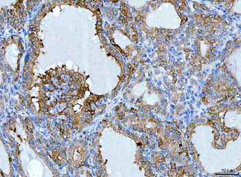
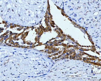
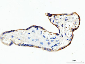
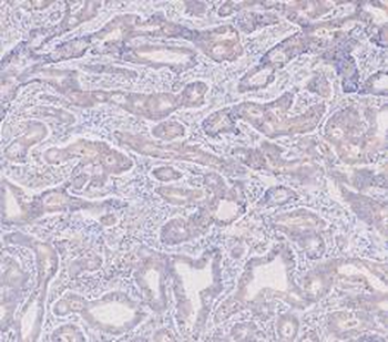
IHC analysis of KRT7 using anti-KRT7 antibody. KRT7 was detected in a paraffin-embedded section of human breast cancer tissue. Heat mediated antigen retrieval was performed in EDTA buffer (pH8.0, epitope retrieval solution). The tissue section was blocked with 10% goat serum. The tissue section was then incubated with 2 µg/ml rabbit anti-KRT7 Antibody overnight at 4C. Peroxidase Conjugated Goat Anti-rabbit IgG was used as secondary antibody and incubated for 30 minutes at 37C. The tissue section was developed using HRP Conjugated Rabbit IgG Super Vision Assay Kit with DAB as the chromogen.
IHC analysis of KRT7 using anti-KRT7 antibody. KRT7 was detected in a paraffin-embedded section of human lung cancer tissue. Heat mediated antige
* Mehrwertsteuer und Versandkosten nicht enthalten. Irrtümer und Preisänderungen vorbehalten