ACE2 antibody was raised against a synthetic peptide corresponding to amino acids near the center of human ACE2.The immunogen is located within amino acids 150 - 200 of ACE2.
ACE2 Antibody is supplied in PBS containing 0.02% sodium azide.
Formulierung:
Liquid
Target-Kategorie:
ACE2
Anwendungsbeschreibung:
Application Notes: WB: 4 µg/mL, IHC: 2 µg/mL, IF: 20 µg/mL.Antibody validated: Western Blot in human, mouse and rat samples, Immunohistochemistry in human, mouse and rat samples, Immunofluorescence in human, mouse, and rat samples. All other applications and species not yet tested
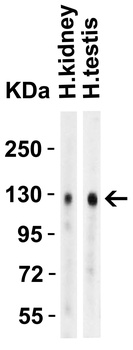
Western Blot Validation in Human Tissues. Loading: 15 µg of lysates per lane. Antibodies: ACE2, orb1239135 (4 µg/mL), 1h incubation at RT in 5% NFDM/TBST. Secondary: Goat anti-rabbit IgG HRP conjugate at 1:10000 dilution.
Immunofluorescence Validation of ACE2 In Caco2 Cells. Immunofluorescent analysis of 4% paraformaldehyde-fixed Caco2 cells labeling ACE2 with orb1239135 at 5 µg/mL, followed by goat anti-rabbit IgG secondary antibody at 1/500 dilution (green) and DAPI staining (blue). Image showing membrane staining on Caco2 cells.
Western Blot Validation in Mouse Stomach Tissue. Loading: 15 µg of lysates per lane. Antibodies: ACE2, orb1239135 (4 µg/mL), 1h incubation at RT in 5% NFDM/TBST. Secondary: Goat anti-rabbit IgG HRP conjugate at 1:10000 dilution.
Western Blot Validation in Rat Brain Tissue. Loading: 15 µg of lysates per lane. Antibodies: ACE2, orb1239135 (4 µg/mL), 1h incubation at RT in 5% NFDM/TBST. Secondary: Goat anti-rabbit IgG HRP conjugate at 1:10000 dilution.
Immunohistochemistry Validation of ACE2 in Human Kidney Tissue. Immunohistochemical analysis of paraffin-embedded human kidney tissue using anti-ACE2 antibody (orb1239135) at 2 µg/ml. Tissue was fixed with formaldehyde and blocked with 10% serum for 1 h at RT, antigen retrieval was by heat mediation with a citrate buffer (pH6). Samples were incubated with primary antibody overnight at 4 C. A goat anti-rabbit IgG H&L (HRP) at 1/250 was used as secondary. Counter stained with Hematoxylin.
Immunofluorescence Validation of ACE2 in Human Kidney Cells. Immunofluorescent analysis of 4% paraformaldehyde-fixed human kidney cells labeling ACE2 with orb1239135 at 20 µg/mL, followed by goat anti-rabbit IgG secondary antibody at 1/500 dilution (green).
Immunofluorescence Validation of ACE2 in Human Testis Tissue. Immunofluorescent analysis of 4% paraformaldehyde-fixed human testis tissue labeling ACE-2 with orb1239135 at 20 µg/mL, followed by goat anti-rabbit IgG secondary antibody at 1/500 dilution (green) and DAPI staining (blue).
Immunofluorescence Validation of ACE2 in Human Lung Tissue. Immunofluorescent analysis of 4% paraformaldehyde-fixed human lung tissue labeling ACE-2 with orb1239135 at 20 µg/mL, followed by goat anti-rabbit IgG secondary antibody at 1/500 dilution (green) and DAPI staining (blue).
Immunofluorescence Validation of ACE2 in Mouse Lung Tissue. Immunofluorescent analysis of 4% paraformaldehyde-fixed mouse lung tissue labeling ACE-2 with orb1239135 at 20 µg/mL, followed by goat anti-rabbit IgG secondary antibody at 1/500 dilution (green) and DAPI staining (blue).
Immunofluorescence Validation of ACE2 in Rat Lung Tissue. Immunofluorescent analysis of 4% paraformaldehyde-fixed rat lung tissue labeling ACE-2 with orb1239135 at 20 µg/mL, followed by goat anti-rabbit IgG secondary antibody at 1/500 dilution (green) and DAPI staining (blue).
* Mehrwertsteuer und Versandkosten nicht enthalten. Irrtümer und Preisänderungen vorbehalten