Anti-hRIP3 antibody (orb1239442) was raised against a peptide corresponding to 18 amino acids near the carboxy terminus of human hRIP3. The immunogen is located within the last 50 amino acids of hRIP3.
Konjugation:
Unconjugated
Alternative Synonym:
hRIP3 Antibody: Rip3, AW107945, 2610528K09Rik, RIP-like protein kinase 3, RIP-3
hRIP3 Antibody is supplied in PBS containing 0.02% sodium azide.
Formulierung:
Liquid
Target-Kategorie:
Ripk3
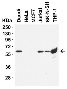
Western Blot Validation in Human Cell Lines. Loading: 15 µg of lysates per lane. Antibodies: hRIP3, orb1239442 (0.5 µg/mL), 1h incubation at RT in 5% NFDM/TBST. Secondary: Goat anti-rabbit IgG HRP conjugate at 1:10000 dilution.
Western Blot Validation in Human Tissues. Loading: 15 µg of lysates per lane. Antibodies: hRIP3, orb1239442 (1 µg/mL), 1h incubation at RT in 5% NFDM/TBST. Secondary: Goat anti-rabbit IgG HRP conjugate at 1:10000 dilution.
Western Blot Validation of hRIP3 in Human THP-1 Cell Lysate. Loading: 15 µg of lysates per lane. Antibodies: hRIP3, orb1239442 (1 µg/mL), 1h incubation at RT in 5% NFDM/TBST. Secondary: Goat anti-rabbit IgG HRP conjugate at 1:10000 dilution. Lane 1-2: human THP-1 cell lysate in the absence (Lane 1) or the presence (Lane 2) of blocking peptide.
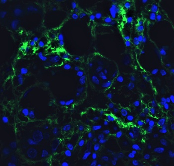
Immunofluorescence Validation of hRIP3 in Molt4 Cells. Immunofluorescent analysis of 4% paraformaldehyde-fixed Molt4 cells labeling hRIP3 with orb1239442 at 20 µg/mL, followed by goat anti-rabbit IgG secondary antibody at 1/500 dilution (green) and DAPI staining (blue).
Immunofluorescence Validation of hRIP3 in Human Breast Tissue. Immunofluorescent analysis of 4% paraformaldehyde-fixed human breast tissue labeling hRIP3 with orb1239442 at 20 µg/mL, followed by goat anti-rabbit IgG secondary antibody at 1/500 dilution (green) and DAPI staining (blue).
Immunofluorescence Validation of hRIP3 in Human Colon Tissue. Immunofluorescent analysis of 4% paraformaldehyde-fixed human colon tissue labeling hRIP3 with orb1239442 at 20 µg/mL, followed by goat anti-rabbit IgG secondary antibody at 1/500 dilution (green) and DAPI staining (blue).
Immunohistochemistry Validation of hRIP3 in Human Liver Tissue. Immunohistochemical analysis of paraffin-embedded human liver tissue using anti-hRIP3 antibody (orb1239442) at 5 µg/ml. Tissue was fixed with formaldehyde and blocked with 10% serum for 1 h at RT, antigen retrieval was by heat mediation with a citrate buffer (pH6). Samples were incubated with primary antibody overnight at 4C. A goat anti-rabbit IgG H&L (HRP) at 1/250 was used as secondary. Counter stained with Hematoxylin.
Immunohistochemistry Validation of hRIP3 in Human Breast Tissue. Immunohistochemical analysis of paraffin-embedded human breast tissue using anti-hRIP3 antibody (orb1239442) at 5 µg/ml. Tissue was fixed with formaldehyde and blocked with 10% serum for 1 h at RT, antigen retrieval was by heat mediation with a citrate buffer (pH6). Samples were incubated with primary antibody overnight at 4C. A goat anti-rabbit IgG H&L (HRP) at 1/250 was used as secondary. Counter stained with Hematoxylin.
Immunohistochemistry Validation of hRIP3 in Human Colon Tissue. Immunohistochemical analysis of paraffin-embedded human colon tissue using anti-hRIP3 antibody (orb1239442) at 2 µg/ml. Tissue was fixed with formaldehyde and blocked with 10% serum for 1 h at RT, antigen retrieval was by heat mediation with a citrate buffer (pH6). Samples were incubated with primary antibody overnight at 4C. A goat anti-rabbit IgG H&L (HRP) at 1/250 was used as secondary. Counter stained with Hematoxylin.
* Mehrwertsteuer und Versandkosten nicht enthalten. Irrtümer und Preisänderungen vorbehalten