
SARS-CoV-2 (COVID-19) Spike Antibody, Unconjugated, Rabbit, Polyclonal
Artikelnummer:
BYT-ORB1239976
- Bilder (14)


| Artikelname: | SARS-CoV-2 (COVID-19) Spike Antibody, Unconjugated, Rabbit, Polyclonal |
| Artikelnummer: | BYT-ORB1239976 |
| Hersteller Artikelnummer: | orb1239976 |
| Alternativnummer: | BYT-ORB1239976-0.02,BYT-ORB1239976-0.1 |
| Hersteller: | Biorbyt |
| Wirt: | Rabbit |
| Kategorie: | Antikörper |
| Applikation: | ELISA, IF, IHC, WB |
| Spezies Reaktivität: | Virus |
| Immunogen: | Anti-SARS-CoV-2 (COVID-19) Spike antibody (orb1239976) was raised against a peptide corresponding to 20 amino acids near the carboxy terminus of SARS-CoV-2 (COVID-19) Spike glycoprotein. The immunogen is located within the last 50 amino acids of SARS-CoV-2 (COVID-19) Spike protein. |
| Konjugation: | Unconjugated |
| Alternative Synonym: | SARS-CoV-2 (COVID-19) Spike Antibody: Severe acute respiratory syndrome coronavirus 2 (SARS-CoV-2), Spike protein, Surface Glycoprotein, covid-19, sars-cov-2 |
| SARS-CoV-2 (COVID-19) Spike Antibody |
| Anwendungsbeschreibung: | Application Notes: WB: 1 µg/mL, IF: 1 µg/mL. IHC: 0.2 µg/mL, Antibody validated: Immunofluorescence and Western blot in human samples. Immunohisochemistry and immunofuorescence in COVID-19 patient samples. It will detect 4 ng of free peptide at 1 µg/mL. The immunogen for this is within the last 50 aa of the spike protein - a peptide corresponding to 20 amino acids near the carboxy terminus of SARS-CoV-2 (COVID-19) Spike glycoprotein. The Extracellular domain (ECD) is from aa 1 to 1208 (full length 1273aa). Therefore, this antibody detects the transmembrane and cytoplasm domains at the C terminus, but does not detect the ECD (which is the region expressed in many commercially available spike proteins). This antibody can be used for the detection of full length spike protein and spike protein in COVID-19 patient samples. All other applications and species not yet tested |
|
|
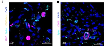
Immunofluorescent Validation of orb1239976 in SARS-CoV-2 Infected Lung Tissue (Singh et al., Nature Microbiology, 2021). Multilabel confocal immunofluorescence microscopy of formalin-fixed paraffin-embedded lung sections from rhesus macaques infected with SARS-CoV-2. SARS-CoV-2 spike-specific antibodies, orb1239976 (k, n) (turquoise) , Ki67 (magenta) and neutrophil marker CD66abce (yellow) (k) , pan-macrophage marker CD68 (magenta) (n) and DAPI (blue). |
|
|
|
|
|
|
|
|
|
|
|
|
|
|
|
|
|
Immunofluorescent Validation of orb1239976 in SARS-CoV-2 Infected Nose and Tonsil (Singh et al., Nature Microbiology, 2021). Multi-label confocal immunofluorescence microscopy of nasal epithelium (20X-b, 63xh) and tonsil (20X-c, 63X-i) from rhesus macaques infected with SARS-CoV-2 with SARS-CoV-2 spike-specific antibodies, orb1239976 (turquoise), DAPI (blue). Rabbit IgG isotype control antibody wasused to stain the tissues to rule out any non-specific staining (e, f). |
|
|
Immunofluorescent Validation of orb1239976 in SARS-CoV-2 Infected Lung Tissue (Singh et al., Nature Microbiology, 2021). Multilabel confocal immunofluorescence microscopy of formalin-fixed paraffin-embedded lung sections from rhesus macaques infected with SARS-CoV-2. SARS-CoV-2 spike-specific antibodies, orb1239976 (turquoise) , Ki67 (magenta) and neutrophil marker CD66abce (yellow) (k-m) , pan-macrophage marker CD68 (magenta) (n-p) , HLA-DR (magenta) and pDC marker CD123 (yellow) (q-s) and DAPI (blue). |
|
|
Immunohistochemistry Validation of SARS-CoV-2 (COVID-19) Spike in COVID-19 Patient Lung. Immunohistochemical analysis of paraffin-embedded COVID-19 patient lung tissue using anti-SARS-CoV-2 (COVID-19) Spike S2 antibody (orb1239976, 0.5 µg/mL). Tissue was fixed with formaldehyde and blocked with 10% serum for 1 h at RT, antigen retrieval was by heat mediation with a citrate buffer (pH6). Samples were incubated with primary antibody overnight at 4 C. A goat anti-rabbit IgG H&L (HRP) at 1/250 was used as secondary. Counter stained with Hematoxylin. Strong spike protein signal was observed in macrophages and airway epithelium of COVID-19 patient lung, but not in non-COVID-19 patient lung. |
|
|
Immunohistochemistry Validation of SARS-CoV-2 (COVID-19) Spike in COVID-19 Patient Lung. Immunohistochemical analysis of paraffin-embedded COVID-19 patient lung tissue using anti-SARS-CoV-2 (COVID-19) Spike S2 antibody (orb1239976, 0.5 µg/mL). Tissue was fixed with formaldehyde and blocked with 10% serum for 1 h at RT, antigen retrieval was by heat mediation with a citrate buffer (pH6). Samples were incubated with primary antibody overnight at 4 C. A goat anti-rabbit IgG H&L (HRP) at 1/250 was used as secondary. Counter stained with Hematoxylin.Strong spike protein signal was observed in macrophages of COVID-19 patient lung. |
|
|
IHC/IF Validation in COVID-19 Patient Sample. (Nuovo et al., 2020) Detection of SARS-CoV-2 proteins in nasopharyngeal swab cell preparations. B. An intense signal for covid-19 spike protein tested by SARS-CoV-2 spike antibodies (orb1239976) was observed in the glandular cells. F-H. Co-expression of spike detected by spike antibodies (orb1239976, 0.2 µg/mL) and envelope proteins detected by envelope antibodies (orb1239971, 2 µg/mL) of SARS-CoV-2 (F) documented localization of each protein to glandular cells (G, yellow). No signal was seen in oral swabs of positive cases (H). Both the spike and envelope protein detected by anti-spike antibodies (orb1239976) and anti-envelope antibodies (orb1239971) produced a signal in the nasopharyngeal swabs of the three cases and no signal was evident in the nasopharyngeal swabs of the seven controls. |
|
|
IF Validation of SARS-CoV2 Spike in COVID-19 Patient Lung. (Magro et al., 2020) SARS-CoV2 spike protein (red, panel C) detected detected by anti-spike antibodies (orb1239976, 0.2 µg/mL) colocalized with C4d (green in panel d, merged in yellow). Spike protein (red, panel g) was also colocalized with C5b-9 (green in panel f&h, merged in yellow). |
|
|
IF Validation of SARS-CoV2 Spike in C |
|
|
Produktgarantie und fachkundiger Support
