SQSTM1 antibody was raised against a 13 amino acid synthetic peptide from near the carboxy terminus of human SQSTM1.The immunogen is located within the last 50 amino acids of SQSTM1.
Konjugation:
Unconjugated
Alternative Synonym:
SQSTM1 Antibody: p60, p62, A170, OSIL, PDB3, ZIP3, p62B, ORCA, Sequestosome-1, EBI3-associated protein of 60 kDa, EBIAP
SQSTM1 Antibody is supplied in PBS containing 0.02% sodium azide.
Formulierung:
Liquid
Target-Kategorie:
SQSTM1
Anwendungsbeschreibung:
Application Notes: WB: 0.5-2 µg/mL, IF: 20 µg/mL, IHC: 2-5 µg/mL,.Antibody validated: Western Blot in human and mouse samples, Immunofluorescence and Immunohistochemistry in human, mouse and rat samples. All other applications and species not yet tested
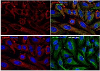
Immunofluorescence Validation of SQSTM1 In HeLa Cells. Immunofluorescent analysis of PFA-fixed HeLa cells labeling SQSTM1 with orb1240011 at 20 µg/mL, followed by goat anti-rabbit IgG secondary antibody at 1/1000 dilution (red) and Hoechst staining (blue). Alpha tubulin was stained with anti-alpha tubulin antibody following by goat anti-mouse IgG secondary antibody (green). Images were captured with confocal microscopy.
Western Blot Validation in Rat Tissues. Loading: 15 µg of lysates per lane. Antibodies: SQSTM1 orb1240011 (1 µg/mL), 1h incubation at RT in 5% NFDM/TBST. Secondary: Goat anti-rabbit IgG HRP conjugate at 1:10000 dilution.
Immunofluorescence Validation of SQSTM1 in Mouse Spleen Tissue. Immunofluorescent analysis of 4% paraformaldehyde-fixed Mouse Spleen Tissue labeling SQSTM1 with orb1240011 at 20 µg/mL, followed by goat anti-rabbit IgG secondary antibody at 1/500 dilution (green) and DAPI staining (blue).
Immunohistochemistry Validation of SQSTM1 in Human Spleen Tissue. Immunohistochemical analysis of paraffin-emb edded Human Spleen Tissue using anti-SQSTM1 antibody (orb1240011) at 2 µg/mL. Tissue was fixed with formaldehyde and blocked with 10% serum for 1 h at RT, antigen retrieval was by heat mediation with a citrate buffer (pH6). Samples were incubated with primary antibody overnight at 4C. A goat anti-rabbit IgG H&L (HRP) at 1/250 was used as secondary. Counter stained with Hematoxylin.
Immunohistochemistry Validation of SQSTM1 in Mouse Spleen Tissue. Immunohistochemical analysis of paraffin-embedded Mouse Spleen Tissue using anti-SQSTM1 antibody (orb1240011) at 2 µg/mL. Tissue was fixed with formaldehyde and blocked with 10% serum for 1 h at RT, antigen retrieval was by heat mediation with a citrate buffer (pH6). Samples were incubated with primary antibody overnight at 4C. A goat anti-rabbit IgG H&L (HRP) at 1/250 was used as secondary. Counter stained with Hematoxylin.
Immunohistochemistry Validation of SQSTM1 in Rat Spleen Tissue. Immunohistochemical analysis of paraffin-embedded Rat Spleen Tissue using anti-SQSTM1 antibody (orb1240011) at 2 µg/mL. Tissue was fixed with formaldehyde and blocked with 10% serum for 1 h at RT, antigen retrieval was by heat mediation with a citrate buffer (pH6). Samples were incubated with primary antibody overnight at 4C. A goat anti-rabbit IgG H&L (HRP) at 1/250 was used as secondary. Counter stained with Hematoxylin.
Induced Expression Validation in Mouse Macrophage Cells. Loading: 15 µg of lysates per lane. Antibodies: SQSTM1 orb1240011 (0.5 µg/mL), 1h incubation at RT in 5% NFDM/TBST. Secondary: Goat anti-rabbit IgG HRP conjugate at 1:10000 dilution. Raw 264.7 cells were treated with LPS (0.3 µg /mL) for different time period (0-24 hrs). There was an increase in SQSTM1 protein expression overtime after LPS treatment.
KO Validation in HEK293T Cells. Loading: 10 µg of HEK293T WT cell lysates or SQSTM1 KO cell lysates. Antibodies: SQSTM1 orb1240011 (1 µg/mL) and beta-actin orb1240312 (1 µg/mL), 1 h incubation at RT in 5% NFDM/TBST. Secondary: Goat Anti-Rabbit IgG HRP conjugate at 1:10000 dilution.
Western Blot Validation in Cell Lines. Loading: 15 µg of lysates per lane. Antibodies: SQSTM1 orb1240011 (0.5 µg/mL), 1 h incubation at RT in 5% NFDM/TBST. Secondary: Goat anti-rabbit IgG HRP conjugate at 1:10000 dilution.
Western Blot Validation in Mouse Tissues. Loading: 15 µg of lysates per lane. Antibodies: SQSTM1 orb1240011 (1 µg/mL), 1h incubation at RT in 5% NFDM/TBST. Secondary: Goat anti-rabbit IgG HRP conjugate at 1:10000 dilution.
* Mehrwertsteuer und Versandkosten nicht enthalten. Irrtümer und Preisänderungen vorbehalten