3-(4-hydroxy-3-nitrophenylacetamido) propionic acid-bovine serum albumin
Konjugation:
Biotin
Alternative Synonym:
Nitrotyrosine, Nitro tyrosine, 3-Nitrotyrosine
Mouse monoclonal to Nitrotyrosine (Biotin). Protein tyrosine nitration results in a post-translational modification that is increasingly receiving attention as an important component of nitric oxide signaling. While multiple nonenzymatic mechanisms are known to be capable of producing nitrated tyrosine residues, most tyrosine nitration events involve catalysis by metalloproteins such as myeloperoxidase, eosino-philperoxidase, myoglobin, the cytochrome P-450s, superoxide dismutase and prostacyclin synthase. Nitrotyrosine may also serve as a biomarker for the effects of reactive nitrogen oxides, based on tyrosine residues becoming nitrated in proteins at sites of inflammation induced tissue injury. The presence of nitro tyrosine-containing proteins therefore has shown high correlation to disease states such as atherosclerosis, Alzheimers disease, Parkinsons disease and amyotrophic lateral sclerosis..
Klonalität:
Monoclonal
Konzentration:
1 mg/ml
Klon-Bezeichnung:
[39B6]
Isotyp:
IgG2a
Puffer:
136.36mM Ethanolamine, 133.23 mM Chlorides, 9.55mM Phosphates, 9.55mM Sodium Bicarbonate
Target-Kategorie:
Nitrotyrosine
Application Verdünnung:
WB (1:1400), IHC (1:100)
Anwendungsbeschreibung:
Application Notes: 0.7 µg/ml of SMC-154 was sufficient for detection of 5 µg SIN-1 treated BSA by Western Blot analysis using Goat anti-mouse IgG:HRP as the secondary antibody
Immunohistochemistry analysis using Mouse Anti-Nitrotyrosine Monoclonal Antibody, Clone 39B6. Tissue: inflamed colon. Species: Mouse. Fixation: Formalin. Primary Antibody: Mouse Anti-Nitrotyrosine Monoclonal Antibody at 1:1000000 for 12 hours at 4C. Secondary Antibody: Biotin Goat Anti-Mouse at 1:2000 for 1 hour at RT. Counterstain: Mayer Hematoxylin (purple/blue) nuclear stain at 200 µl for 2 minutes at RT. Magnification: 40x. With anti-microbial.
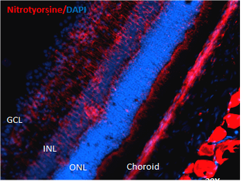
Immunohistochemistry analysis using Mouse Anti-Nitrotyrosine Monoclonal Antibody, Clone 39B6. Tissue: liver tissue. Species: Rat. Primary Antibody: Mouse Anti-Nitrotyrosine Monoclonal Antibody at 1:1000. Secondary Antibody: FITC Goat Anti-Mouse (green).
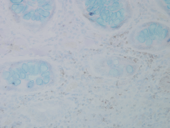
Immunohistochemistry analysis using Mouse Anti-Nitrotyrosine Monoclonal Antibody, Clone 39B6. Tissue: colon carcinoma. Species: Human. Fixation: Formalin. Primary Antibody: Mouse Anti-Nitrotyrosine Monoclonal Antibody at 1:25000 for 12 hours at 4C. Secondary Antibody: Biotin Goat Anti-Mouse at 1:2000 for 1 hour at RT. Counterstain: Mayer Hematoxylin (purple/blue) nuclear stain at 200 µl for 2 minutes at RT. Magnification: 40x.
Western Blot analysis of Human A549 cells showing detection of Multiple Bands Nitrotyrosine protein using Mouse Anti-Nitrotyrosine Monoclonal Antibody, Clone 39B6. Lane 1: MW ladder. Lane 2: Human A549 Cells 15 ug). Load: 15 ug. Block: 5% Skim Milk Powder in TBST. Primary Antibody: Mouse Anti-Nitrotyrosine Monoclonal Antibody at 1:1000 for 2.5 hours at RT with shaking. Secondary Antibody: Goat anti-mouse IgG:HRP at 1:1000 for 1 hour at RT with shaking. Color Development: Chemiluminescent for HRP (Moss) for 5 min in RT. Predicted/Observed Size: Multiple Bands.
Western Blot analysis of Human Recombinant Protein showing detection of Multiple Bands Nitrotyrosine protein using Mouse Anti-Nitrotyrosine Monoclonal Antibody, Clone 39B6. Lane 1: MW Ladder. Lane 2: hASYN Monomer (3.84 ug). Lane 3: Nitrosylated hASYN (3.84 ug). Block: 5% Skim Milk Powder in TBST. Primary Antibody: Mouse Anti-Nitrotyrosine Monoclonal Antibody at 1:1000 for 2 hours at RT with shaking. Secondary Antibody: Goat anti-mouse IgG:HRP at 1:4000 for 2 hour at RT with shaking. Color Development: Chemiluminescent for HRP (Moss) for 5 min in RT. Predicted/Observed Size: Multiple Bands.
Western Blot analysis of Human HEK293 cells showing detection of Nitrotyrosine protein using Mouse Anti-Nitrotyrosine Monoclonal Antibody, Clone 39B6. Lane 1: MW Ladder. Lane A: Nitrosylated-HEK293 (15uL). Lane B: HEK293 (15 ug). Block: 5% Skim Milk Powder in TBST. Primary Antibody: Mouse Anti-Nitrotyrosine Monoclonal Antibody diluted in 1.5% BSA and TBST for 1 hours at RT with shaking. Secondary Antibody: Goat anti-mouse IgG: HRP at 1:4000 for 1 hour at RT with shaking. Predicted/Observed Size: Multiple Bands.
Immunohistochemistry analysis using Mouse Anti-Nitrotyrosine Monoclonal Antibody, Clone 39B6. Tissue: backskin. Species: Mouse. Fixation: Bouins Fixative and paraffin-embedded. Primary Antibody: Mouse Anti-Nitrotyrosine Monoclonal Antibody at 1:100 for 1 hour at RT. Secondary Antibody: FITC Goat Anti-Mouse (green) at 1:50 for 1 hour at RT. Backskin obtained from transgenic mice.
* Mehrwertsteuer und Versandkosten nicht enthalten. Irrtümer und Preisänderungen vorbehalten