Mouse monoclonal to CaVbeta2 (Biotin). Cav Beat subunits are involved in the transport of the pore forming alpha1 subunit to the plasma membrane. They also shield an ER Retention signal on the alpha1 subunit, thereby guiding the pore-forming subunit to the target membrane. They also determine the biophysical properties of the calcium channel..
136.36mM Ethanolamine, 133.23 mM Chlorides, 9.55mM Phosphates, 9.55mM Sodium Bicarbonate
Target-Kategorie:
CavBeta2
Application Verdünnung:
WB (1:1000), IHC (1:1000), ICC/IF (1:100)
Anwendungsbeschreibung:
Application Notes: 1 µg/ml of SMC-332 was sufficient for detection of Cavbeta2 in 10 µg of rat brain lysate by colorimetric immunoblot analysis using Goat anti-mouse IgG:HRP as the secondary antibody
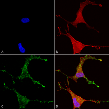
Immunocytochemistry/Immunofluorescence analysis using Mouse Anti-Cav beta 2 Monoclonal Antibody, Clone S8B-1. Tissue: Neuroblastoma cells (SH-SY5Y). Species: Human. Fixation: 4% PFA for 15 min. Primary Antibody: Mouse Anti-Cav beta 2 Monoclonal Antibody at 1:50 for overnight at 4C with slow rocking. Secondary Antibody: AlexaFluor 488 at 1:1000 for 1 hour at RT. Counterstain: Phalloidin-iFluor 647 (red) F-Actin stain, Hoechst (blue) nuclear stain at 1:800, 1.6mM for 20 min at RT. (A) Hoechst (blue) nuclear stain. (B) Phalloidin-iFluor 647 (red) F-Actin stain. (C) Cav beta 2 Antibody (D) Composite.
Immunohistochemistry analysis using Mouse Anti-Cav Beta2 Calcium Channel Monoclonal Antibody, Clone S8b-1. Tissue: backskin. Species: Mouse. Fixation: Bouins Fixative and paraffin-embedded. Primary Antibody: Mouse Anti-Cav Beta2 Calcium Channel Monoclonal Antibody at 1:100 for 1 hour at RT. Secondary Antibody: FITC Goat Anti-Mouse (green) at 1:50 for 1 hour at RT. Localization: All nuclei. Some nuclei also staining higher up in epidermis.
Western Blot analysis of Human Cell line lysates showing detection of Cav Beta2 Calcium Channel protein using Mouse Anti-Cav Beta2 Calcium Channel Monoclonal Antibody, Clone S8b-1. Load: 15 µg. Block: 1.5% BSA for 30 minutes at RT. Primary Antibody: Mouse Anti-Cav Beta2 Calcium Channel Monoclonal Antibody at 1:1000 for 2 hours at RT. Secondary Antibody: Sheep Anti-Mouse IgG: HRP for 1 hour at RT.
Immunocytochemistry/Immunofluorescence analysis using Mouse Anti-Cav Beta2 Calcium Channel Monoclonal Antibody, Clone S8b-1. Tissue: HaCaT cells. Species: Human. Fixation: Cold 100% methanol for 10 minutes at -20C. Primary Antibody: Mouse Anti-Cav Beta2 Calcium Channel Monoclonal Antibody at 1:100 for 1 hour at RT. Secondary Antibody: FITC Goat Anti-Mouse (green) at 1:50 for 1 hour at RT. Localization: All cells positive. Bright dottiness located throughout cytoplasm and in nuclei.
* Mehrwertsteuer und Versandkosten nicht enthalten. Irrtümer und Preisänderungen vorbehalten