TRPC7 Antibody (Biotin), IgG1, Clone: [N64A/36 Formerly sold as S64A-36], Mouse, Monoclonal
Artikelnummer:
BYT-ORB148858
Hersteller Artikelnummer:
orb148858
Alternativnummer:
BYT-ORB148858-100
Hersteller:
Biorbyt
Wirt:
Mouse
Kategorie:
Antikörper
Applikation:
AM, ICC, IF, IHC, IP, WB
Spezies Reaktivität:
Human, Mouse, Rat
Immunogen:
Synthetic peptide amino acids 845-862 of human TRPC7
Konjugation:
Biotin
Alternative Synonym:
TRP7, KNP3, TRPM2, transient receptor potential cation channel subfamily C member 7
Mouse monoclonal to TrpC7 (Biotin). Transient receptor potential cation channel, subfamily C, member 7, also known as TRPC7, is a non-selective cation channel that is directly activated by DAG. TrpC7 shows constitutive activity and susceptibility to negative regulation by extracellular Ca2+. Because of this, TrpC7 plays an important role in the Ca2+ signaling pathway. TrpC7 is also expressed abundantly in the heart, and combined with its ability to act as a Ca2+ channel, TrpC7 might contribute to the process of heart failure..
136.36mM Ethanolamine, 133.23 mM Chlorides, 9.55mM Phosphates, 9.55mM Sodium Bicarbonate
Target-Kategorie:
TRPC7
Application Verdünnung:
WB (1:1000), IHC (1:1000), ICC/IF (1:100)
Anwendungsbeschreibung:
Application Notes: 1 µg/ml of SMC-343 was sufficient for detection of TrpC7 in 10 µg of rat brain lysate by colorimetric immunoblot analysis using goat anti-mouse IgG:HRP as the secondary antibody
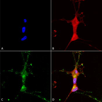
Immunocytochemistry/Immunofluorescence analysis using Mouse Anti-TrpC7 Monoclonal Antibody, Clone N64A/36. Tissue: Neuroblastoma cells (SH-SY5Y). Species: Human. Fixation: 4% PFA for 15 min. Primary Antibody: Mouse Anti-TrpC7 Monoclonal Antibody at 1:100 for overnight at 4C with slow rocking. Secondary Antibody: AlexaFluor 488 at 1:1000 for 1 hour at RT. Counterstain: Phalloidin-iFluor 647 (red) F-Actin stain, Hoechst (blue) nuclear stain at 1:800, 1.6mM for 20 min at RT. (A) Hoechst (blue) nuclear stain. (B) Phalloidin-iFluor 647 (red) F-Actin stain. (C) TrpC7 Antibody (D) Composite.
Immunohistochemistry analysis using Mouse Anti-TrpC7 Monoclonal Antibody, Clone N64A/36. Tissue: backskin. Species: Mouse. Fixation: Bouins Fixative and paraffin-embedded. Primary Antibody: Mouse Anti-TrpC7 Monoclonal Antibody at 1:100 for 1 hour at RT. Secondary Antibody: FITC Goat Anti-Mouse (green) at 1:50 for 1 hour at RT. Localization: Everything.
Western Blot analysis of Rat brain membrane lysate showing detection of TrpC7 protein using Mouse Anti-TrpC7 Monoclonal Antibody, Clone N64A/36. Load: 15 µg. Block: 1.5% BSA for 30 minutes at RT. Primary Antibody: Mouse Anti-TrpC7 Monoclonal Antibody at 1:1000 for 2 hours at RT. Secondary Antibody: Sheep Anti-Mouse IgG: HRP for 1 hour at RT.
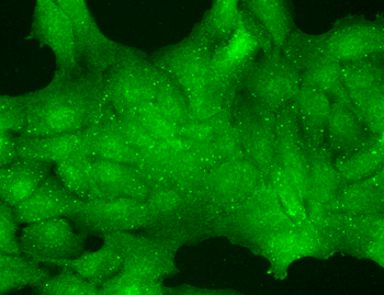
Immunocytochemistry/Immunofluorescence analysis using Mouse Anti-TrpC7 Monoclonal Antibody, Clone N64A/36. Tissue: HaCaT cells. Species: Human. Fixation: Cold 100% methanol for 10 minutes at -20C. Primary Antibody: Mouse Anti-TrpC7 Monoclonal Antibody at 1:100 for 1 hour at RT. Secondary Antibody: FITC Goat Anti-Mouse (green) at 1:50 for 1 hour at RT. Localization: Nuclear staining.
* Mehrwertsteuer und Versandkosten nicht enthalten. Irrtümer und Preisänderungen vorbehalten