Bovine, Canine, Gallus, Hamster, Human, Mouse, Rabbit, Rat
Immunogen:
Human HSP60 produced through recombinant DNA methods in E.coli
Konjugation:
Biotin
Alternative Synonym:
HSPD1, HSP60, 60 kDa heat shock protein, mitochondrial, Chaperonin 60, CPN60, HuCHA60, Heat shock protein family D member 1, GroEL homolog, mitochondrial, GROEL, HLD4, HSP 60, HSP65, SPG 13
Rabbit polyclonal to Hsp60 (Biotin). In both prokaryotic and eukaryotic cells, the misfolding In both prokaryotic and eukaryotic cells, the misfolding...
136.36mM Ethanolamine, 133.23 mM Chlorides, 9.55mM Phosphates, 9.55mM Sodium Bicarbonate.
Target-Kategorie:
HSP60
Application Verdünnung:
WB (1:1000), ICC/IF (1:100)
Anwendungsbeschreibung:
Application Notes: 1 µg/ml of SPC-105 was sufficient for detection of HSP60 in 20 µg of heat shocked HeLa cell lysate by colorimetric immunoblot analysis using goat anti-mouse IgG as the secondary antibody
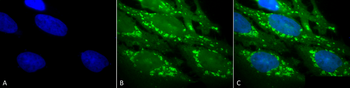
Immunocytochemistry/Immunofluorescence analysis using Rabbit Anti-Hsp60 Polyclonal Antibody. Tissue: Heat Shocked Cervical cancer cell line (HeLa). Species: Human. Fixation: 2% Formaldehyde for 20 min at RT. Primary Antibody: Rabbit Anti-Hsp60 Polyclonal Antibody at 1:100 for 12 hours at 4C. Secondary Antibody: FITC Goat Anti-Rabbit (green) at 1:200 for 2 hours at RT. Counterstain: DAPI (blue) nuclear stain at 1:40000 for 2 hours at RT. Localization: Mitochondrion matrix. Magnification: 100x. (A) DAPI (blue) nuclear stain. (B) Anti-Hsp60 Antibody. (C) Composite. Heat Shocked at 42C for 1h.
Western blot analysis of Human, Dog, Mouse SKBR3, MDCK, and MEF cell line lysates showing detection of HSP60 protein using Rabbit Anti-HSP60 Polyclonal Antibody. Lane 1: Recom. Human Hsp60 (100ng), Lane2, 3 and 4: SKBR3 lysate (human), MDCK lysate (dog) and MEF lysate (mouse) (al at 7.5ug). Primary Antibody: Rabbit Anti-HSP60 Polyclonal Antibody at 1:1000.
Immunocytochemistry/Immunofluorescence analysis using Rabbit Anti-Hsp60 Polyclonal Antibody. Tissue: Heat Shocked Cervical cancer cell line (HeLa). Species: Human. Fixation: 2% Formaldehyde for 20 min at RT. Primary Antibody: Rabbit Anti-Hsp60 Polyclonal Antibody at 1:100 for 12 hours at 4C. Secondary Antibody: APC Goat Anti-Rabbit (red) at 1:200 for 2 hours at RT. Counterstain: DAPI (blue) nuclear stain at 1:40000 for 2 hours at RT. Localization: Mitochondrion matrix. Magnification: 20x. (A) DAPI (blue) nuclear stain. (B) Anti-Hsp60 Antibody. (C) Composite. Heat Shocked at 42C for 1h.
* Mehrwertsteuer und Versandkosten nicht enthalten. Irrtümer und Preisänderungen vorbehalten