Human Rab5 synthetic peptide conjugated to KLH, identical to dog Rab5 sequence over the residues
Konjugation:
Biotin
Alternative Synonym:
Rab 5A, RAS associated protein RAB5A, Ras related protein Rab 5 A
Rabbit polyclonal to Rab5 (Biotin). Rab5 is a 24kDa member of the Rab family of small guanosine triphosphatases (GTPases), Ras superfamily. Rab GTPases are central regulators of membrane trafficking in the eukaryotic cell. Their regulatory capacity depends on their ability to cycle between the GDP -bound inactive and GTP-bound active states. This conversion is regulated by GDP/GTP exchange factors (GEPs), GDP dissociation inhibitors (GDIs) and GTPase-activating proteins (GAPs). Activation of a Rab protein is coupled to its association with intracellular membranes, allowing it to recruit downstream effector proteins to the cytoplasmic surface of a subcellular compartment. Through these proteins, Rab GTPases regulate vesicle formation, actin- and tubulin-dependent vesicle movement, and membrane fusion(1). Rab proteins contain conserved regions involved in guanine-nucleotide binding, and hyper variable COHO-terminal domains with a cysteine motif implicated in subcellular targeting. Post-translational modification of the cysteine motif with one or two geranyl groups is essential for the membrane association and correct intracellular localization of Rab proteins(3). Each Rab shows a characteristic subcellular distribution. In particular, Rab5 is ubiquitously expressed in human tissues. It localizes mainly to early endosomes, but is also present on the plasma membrane. It regulates the fusion between endocytic vesicles and early endosomes, as well as the homotypic fusion between early endosomes. Among the proteins recruited by the GTP-bound active Rab5 are Rabaptin-5 and EEA1. Anti-Rab5 may be used as an early endosome marker....
136.36mM Ethanolamine, 133.23 mM Chlorides, 9.55mM Phosphates, 9.55mM Sodium Bicarbonate.
Target-Kategorie:
Rab5
Application Verdünnung:
WB (1:1000), IHC (1:100), ICC/IF (1:80)
Anwendungsbeschreibung:
Application Notes: 1 µl/ml of SPC-168 was sufficient for detection of Rab5 in 15 µg of HeLa lysate by ECL immunoblot analysis using Donkey anti-rabbit IgG:HRP as the secondary antibody
Immunocytochemistry/Immunofluorescence analysis using Rabbit Anti-Rab5 Polyclonal Antibody. Tissue: Cervical cancer cell line (HeLa). Species: Human. Fixation: 2% Formaldehyde for 20 min at RT. Primary Antibody: Rabbit Anti-Rab5 Polyclonal Antibody at 1:80 for 12 hours at 4C. Secondary Antibody: R-PE Goat Anti-Rabbit (yellow) at 1:200 for 2 hours at RT. Counterstain: DAPI (blue) nuclear stain at 1:40000 for 2 hours at RT. Localization: Cytoplasm. Melanosome. Nucleus. Magnification: 100x. (A) DAPI (blue) nuclear stain. (B) Anti-Rab5 Antibody. (C) Composite.
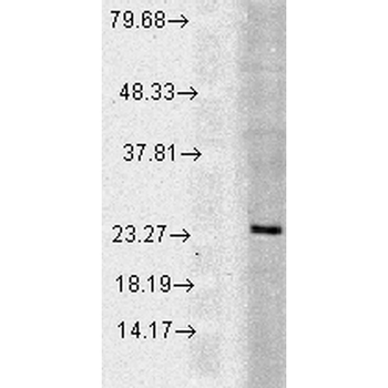
Western blot analysis of Human Cell line lysates showing detection of Rab5 protein using Rabbit Anti-Rab5 Polyclonal Antibody. Load: 15 µgprotein. Block: 1.5% BSA for 30 minutes at RT. Primary Antibody: Rabbit Anti-Rab5 Polyclonal Antibody at 1:1000 for 2 hours at RT. Secondary Antibody: Donkey Anti-Rabbit IgG: HRP for 1 hour at RT.
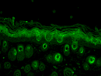
Immunohistochemistry analysis using Rabbit Anti-Rab5 Polyclonal Antibody. Tissue: backskin. Species: Mouse. Fixation: Bouins Fixative Solution. Primary Antibody: Rabbit Anti-Rab5 Polyclonal Antibody at 1:100 for 1 hour at RT. Secondary Antibody: FITC Goat Anti-Rabbit (green) at 1:50 for 1 hour at RT. Localization: Cytoplasm.
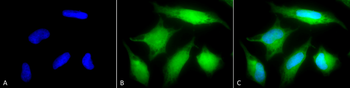
Immunocytochemistry/Immunofluorescence analysis using Rabbit Anti-Rab5 Polyclonal Antibody. Tissue: Cervical cancer cell line (HeLa). Species: Human. Fixation: 2% Formaldehyde for 20 min at RT. Primary Antibody: Rabbit Anti-Rab5 Polyclonal Antibody at 1:80 for 12 hours at 4C. Secondary Antibody: FITC Goat Anti-Rabbit (green) at 1:200 for 2 hours at RT. Counterstain: DAPI (blue) nuclear stain at 1:40000 for 2 hours at RT. Localization: Cytoplasm. Melanosome. Nucleus. Magnification: 20x. (A) DAPI (blue) nuclear stain. (B) Anti-Rab5 Antibody. (C) Composite.
* Mehrwertsteuer und Versandkosten nicht enthalten. Irrtümer und Preisänderungen vorbehalten