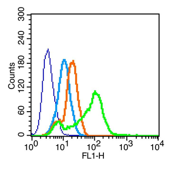
Flow cytometric analysis of mouse spleen cell using DC-SIGN antibody.
Blank control (blue line): MCF7 (blue). Primary Antibody (green line): Rabbit Anti-DC-SIGN antibody, Dilution: 1 µg/10 6 cells, Isotype Control Antibody (orange line): Rabbit IgG. Secondary Antibody (white blue line): F (ab)2 fragment goat anti-rabbit IgG-FITC. Dilution: 1 µg/Test. Protocol, The cells were fixed with 2% paraformaldehyde for 10 min at room temperature. Cells stained with Primary Antibody for 30 min at room temperature. The cells were then incubated in 1X PBS/2% BSA/10% goat serum to block non-specific protein-protein interactions followed by the antibody for 15 min at room temperature. The secondary antibody used for 40 min at room temperature. Acquisition of 20000 events was performed.
Paraformaldehyde-fixed, paraffin embedded (human cervical cancer), Antigen retrieval by boiling in sodium citrate buffer (pH6.0) for 15 min, Block endogenous peroxidase by 3% hydrogen peroxide for 20 minutes, Blocking buffer (normal goat serum) at 37C for 30 min, Antibody incubation with (DC-SIGN) Polyclonal Antibody, Unconjugated (orb156554) at 1:400 overnight at 4C, followed by a conjugated secondary for 20 minutes and DAB staining.
Paraformaldehyde-fixed, paraffin embedded (human liver carcinoma), Antigen retrieval by boiling in sodium citrate buffer (pH6.0) for 15 min, Block endogenous peroxidase by 3% hydrogen peroxide for 20 minutes, Blocking buffer (normal goat serum) at 37C for 30 min, Antibody incubation with (DC-SIGN) Polyclonal Antibody, Unconjugated (orb156554) at 1:400 overnight at 4C, followed by a conjugated secondary for 20 minutes and DAB staining.
Sample: Hela Cell Lysate at 40 ug, Hcclm3 Cell Lysate at 40 ug, Primary: Anti-DC-SIGN (orb156554) at 1/300 dilution, Secondary: IRDye800CW Goat Anti-Rabbit IgG at 1/20000 dilution, Predicted band size: 45 kD, Observed band size: 50 kD.
* Mehrwertsteuer und Versandkosten nicht enthalten. Irrtümer und Preisänderungen vorbehalten