CD antigen CD62P,CD62 antigen-like family member P,CD62P,GMP-140,granule membrane protein 140,Grmp,LECAM3,leukocyte-endothelial cell adhesion molecule 3,PADGEM,platelet activation dependent granule-external membrane protein,P-selectin
Recombinant antibody was purified from cell culture supernatant
Formulierung:
Liquid
Target-Kategorie:
P-Selectin/CD62P
Application Verdünnung:
Flow-Cyt Live or cells fixed in 4% formaldehyde and permeabilized with 90% methanol. 1 µl per 1 x 10 6 cells. Immunocytochemistry (ICC) 1:100 to 1:500. Epitope retrieval with citrate buffer pH 6.0 is recommended for FFPE cell sections. Immunohistochemistr
Anwendungsbeschreibung:
Application Notes: All western blot analysis is performed using 5% Milk-TBST for blocking and as antibody diluent. Primary antibody is incubated overnight
Detection of mouse P-Selectin/CD62P (shaded) in b.END3 cells by flow cytometry. Antibody: Rabbit anti-P-Selectin/CD62P recombinant monoclonal antibody (orb1784574) or isotype control (unshaded). Secondary: DyLight 650-conjugated goat anti-rabbit IgG.
Detection of human P-Selectin/CD62P (shaded) in Hel92.1.7 cells by flow cytometry. Antibody: Rabbit anti-P-Selectin/CD62P recombinant monoclonal antibody (orb1784574) or isotype control (unshaded). Secondary: DyLight 650-conjugated goat anti-rabbit IgG.
Detection of mouse P-Selectin/CD62P by immunocytochemistry. Sample: FFPE section of bEnd.3 cells. Antibody: Rabbit anti-P-Selectin/CD62P recombinant monoclonal antibody (orb1784574). Secondary: HRP-conjugated goat anti-rabbit IgG.
Detection of human P-Selectin/CD62P by immunocytochemistry. Sample: FFPE section of HEL 92.1.7 cells. Antibody: Rabbit anti-P-Selectin/CD62P recombinant monoclonal antibody (orb1784574). Secondary: HRP-conjugated goat anti-rabbit IgG.
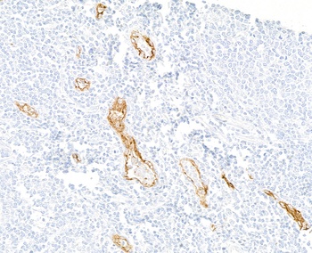
Detection of human P-Selectin/CD62P by immunohistochemistry. Sample: FFPE section of tonsil. Antibody: Rabbit anti-P-Selectin/CD62P recombinant monoclonal antibody (orb1784574). Secondary: HRP-conjugated goat anti-rabbit IgG.
Detection of mouse P-Selectin/CD62P by immunohistochemistry. Sample: FFPE section of mouse ovary. Antibody: Rabbit anti-P-Selectin/CD62P recombinant monoclonal antibody (orb1784574). Secondary: HRP-conjugated goat anti-rabbit IgG.
Detection of human P-Selectin/CD62P by western blot of immunoprecipitates. Samples: Whole cell lysate (1.0 mg per IP reaction, 20% of IP loaded) from HEL 92.1.7 cells prepared using NETN lysis buffer. Antibodies: Rabbit anti-P-Selectin/CD62P recombinant monoclonal antibody (orb1784574) used for IP at 6 µl/mg lysate.
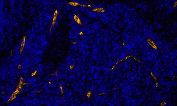
Detection of human P-Selectin/CD62P (orange) by immunofluorescence. Sample: FFPE section of metastatic lymph node. Antibody: Rabbit anti-P-Selectin/CD62P recombinant monoclonal antibody (orb1784574) used at 1:100. Secondary: Invitrogen Goat anti-rabbit IgG Alexa Fluor(TM) Plus 647 1:400. Counterstain: DAPI (blue).
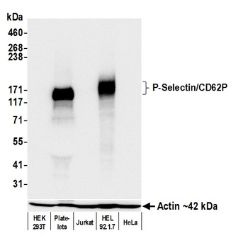
Detection of human P-Selectin/CD62P by western blot. Samples: Whole cell lysate (50 µg) from HEK293T, Platelets, Jurkat, HEL 92.1.7, and HeLa cells prepared using NETN lysis buffer. Antibody: Rabbit anti-P-Selectin/CD62P recombinant monoclonal antibody (orb1784574) used at 1:1000. Secondary: HRP-conjugated goat anti-rabbit IgG. Detection: Chemiluminescence with an exposure time of 10 seconds. Lower Panel: Rabbit anti-Actin recombinant monoclonal antibody.
Detection of mouse P-Selectin/CD62P by western blot. Samples: Whole cell lysate (50 µg) from NIH 3T3, bEnd.3 (10 µg) , CH27, Spleen, and CT26 cells prepared using NETN lysis buffer. Antibody: Rabbit anti-P-Selectin/CD62P recombinant monoclonal antibody (orb1784574) used at 1:1000. Secondary: HRP-conjugated goat anti-rabbit IgG. Detection: Chemiluminescence with an exposure time of 10 seconds. Lower Panel: Rabbit anti-Actin recombinant monoclonal antibody.
* Mehrwertsteuer und Versandkosten nicht enthalten. Irrtümer und Preisänderungen vorbehalten