E.coli-derived human LIN7C recombinant protein (Position: M1-A73).
Konjugation:
Unconjugated
Alternative Synonym:
LIN 7 C, lin 7 homolog C (C. elegans), LIN 7C, LIN7C, MALS 3, MALS3, Mammalian lin seven protein 3, Protein lin 7 homolog C, Veli 3, VELI3, Vertebrate lin 7 homolog 3
Anti-LIN7C Antibody. Tested in ELISA, IHC, WB, Flow Cytometry applications. This antibody reacts with Human, Rat.
Klonalität:
Polyclonal
Konzentration:
Adding 0.2 ml of distilled water will yield a concentration of 500 µg/ml.
Western blot, 0.25-0.5 µg/ml, Human, Rat Immunohistochemistry (Paraffin-embedded Section), 2-5 µg/ml, Human, Rat Flow Cytometry (Fixed), 1-3 µg/1x10 6 cells, Human ELISA, 0.1-0.5 µg/ml, -
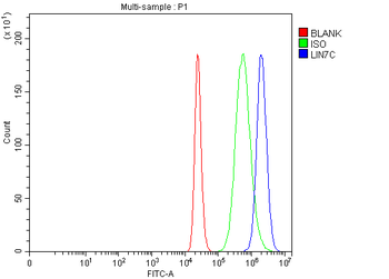
Flow Cytometry analysis of THP-1 cells using anti-LIN7C antibody. Overlay histogram showing THP-1 cells (Blue line). The cells were fixed with 4% paraformaldehyde and blocked with 10% normal goat serum. And then incubated with rabbit anti-LIN7C Antibody (1 µg/1x10 6 cells) for 30 min at 20C. DyLight488 conjugated goat anti-rabbit IgG (5-10 µg/1x10 6 cells) was used as secondary antibody for 30 minutes at 20C. Isotype control antibody (Green line) was rabbit IgG (1 µg/1x10 6) used under the same conditions. Unlabelled sample (Red line) was also used as a control.
Flow Cytometry analysis of THP-1 cells using anti-LIN7C antibody (
Flow Cytometry analysis of THP-1 cells using anti-LIN7C antibody. Overlay histogram showing THP-1 cells (Blue line). The cells were fixed with 4% paraformaldehyde and blocked with 10% normal goat serum. And then incubated with rabbit anti-LIN7C Antibody (1 µg/1x10 6 cells) for 30 min at 20C. DyLight488 conjugated goat anti-rabbit IgG (5-10 µg/1x10 6 cells) was used as secondary antibody for 30 minutes at 20C. Isotype control antibody (Green line) was rabbit IgG (1 µg/1x10 6) used under the same conditions. Unlabelled sample without incubation with primary antibody and secondary antibody (Red line) was used as a blank control.
IHC analysis of LIN7C using anti-LIN7C antibody. LIN7C was detected in a paraffin-embedded section of human adrenal adenomas tissue. Heat mediated antigen retrieval was performed in EDTA buffer (pH8.0, epitope retrieval solution). The tissue section was blocked with 10% goat serum. The tissue section was then incubated with 2 µg/ml rabbit anti-LIN7C Antibody overnight at 4C. Peroxidase Conjugated Goat Anti-rabbit IgG was used as secondary antibody and incubated for 30 minutes at 37C. The tissue section was developed using HRP Conjugated Rabbit IgG Super Vision Assay Kit with DAB as the chromogen.
IHC analysis of LIN7C using anti-LIN7C antibody. LIN7C was detected in a paraffin-embedded section of human bladder urothelial carcinoma tissue. Heat mediated antigen retrieval was performed in EDTA buffer (pH8.0, epitope retrieval solution). The tissue section was blocked with 10% goat serum. The tissue section was then incubated with 2 µg/ml rabbit anti-LIN7C Antibody overnight at 4C. Peroxidase Conjugated Goat Anti-rabbit IgG was used as secondary antibody and incubated for 30 minutes at 37C. The tissue section was developed using HRP Conjugated Rabbit IgG Super Vision Assay Kit with DAB as the chromogen.
IHC analysis of LIN7C using anti-LIN7C antibody. LIN7C was detected in a paraffin-embedded section of human colorectal adenocarcinoma tissue. Heat mediated antigen retrieval was performed in EDTA buffer (pH8.0, epitope retrieval solution). The tissue section was blocked with 10% goat serum. The tissue section was then incubated with 2 µg/ml rabbit anti-LIN7C Antibody overnight at 4C. Peroxidase Conjugated Goat Anti-rabbit IgG was used as secondary antibody and incubated for 30 minutes at 37C. The tissue section was developed using HRP Conjugated Rabbit IgG Super Vision Assay Kit with DAB as the chromogen.
IHC analysis of LIN7C using anti-LIN7C antibody. LIN7C was detected in a paraffin-embedded section of rat kidney tissue. Heat mediated antigen retrieval was performed in EDTA buffer (pH8.0, epitope retrieval solution). The tissue section was blocked with 10% goat serum. The tissue section was then incubated with 2 µg/ml rabbit anti-LIN7C Antibody overnight at 4C. Peroxidase Conjugated Goat Anti-rabbit IgG was used as secondary antibody and incubated for 30 minutes at 37C. The tissue section was developed using HRP Conjugated Rabbit IgG Super Vision Assay Kit with DAB as the chromogen.
Western blot analysis of LIN7C using anti-LIN7C antibody. Electrophoresis was performed on a 5-20% SDS-PAGE gel at 70V (Stacking gel) / 90V (Resolving gel) for 2-3 hours. The sample well of each lane was loaded with 30 ug of sample under reducing conditions. Lane 1: human RT4 whole cell lysates, Lane 2: rat brain tissue lysates. After electrophoresis, pro
* Mehrwertsteuer und Versandkosten nicht enthalten. Irrtümer und Preisänderungen vorbehalten