Biological Origin: Human. Biological Activity: Thioflavin T curve shows less beta-sheet aggregation when Type 2 monomers are seeded with PFFs compared to Type 1 monomers seeded with PFFs. Application Notes: Certified 95% pure using SDS-PAGE analysis. Low endotoxin <5 EU/mL 2mg/mL
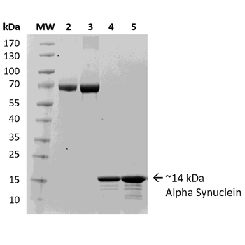
SDS-PAGE of ~14 kDa Human Recombinant Alpha Synuclein Protein Monomer. Lane 1: Molecular Weight Ladder (MW). Lane 2: BSA (2.5 µg). Lane 3: BSA (5 µg). Lane 4: Alpha Synuclein Protein Monomer (2.5 µg). Lane 5: Alpha Synuclein Protein Monomer (5 µg).
Thioflavin T is a fluorescent dye that binds to beta sheet-rich structures, such as those in alpha synuclein fibrils. Upon binding, the emission spectrum of the dye experiences a red-shift and increased fluorescence intensity. Thioflavin T emission curves show increased fluorescence (correlated to alpha synuclein protein aggregation) over time when 10 µM of Type 1 alpha synuclein pre-formed fibrils is combined with 100 µM of Type 1 alpha synuclein monomer, compared to 10 µM of Type 1 alpha synuclein pre-formed fibrils combined with 100 µM Type 2 alpha synuclein monomer. Type 2 fibrils do not seed type 2 monomers (data not shown). Thioflavin T ex = 450 nm, em = 485 nm.
Type 2 monomers are currently undergoing testing in a Real-Time Quaking-Induced Conversion (RT-QuIC) assay. At 10 µg/well there was discrimination between positive and negative CSF samples, and the unseeded reaction occurred later than either the LB BH or the positive CSF sample. This suggests Type 2 monomers could potentially be used as a substrate for alpha synuclein RT-QuIC. Further testing/optimization is underway. LB BH: 10% Lewy body disease, CSF +: CSF from patient with neuropathologically confirmed alpha-synucleinopathy, CSF -: CSF from patient with no evidence of alpha-synuclein deposition at postmortem. Image source: Alison Green, Graham Fairfoul
Evaluation of a-syn toxicity on primary mouse cortical neurons. Mitochondrial dehydrogenase activity reduces yellow MTT to dark blue formazan crystals, a reaction catalyzed in living cells. Cell viability was assessed with an MTT assay and displayed as % of vehicle control (VC). Data are presented as bar graphs and standard deviation.
Evaluation of a-syn toxicity on primary mouse cortical neurons. Lactate dehydrogenase (LDH) is a soluble enzyme present in the cytosol that is released upon cell death. Toxicity was assessed with an LDH assay and displayed as % of vehicle control (VC). Data are presented as bar graphs and standard deviation. For statistical analysis One-way ANOVA followed by Bonferroni post-hoc test (vs VC) was used.
* Mehrwertsteuer und Versandkosten nicht enthalten. Irrtümer und Preisänderungen vorbehalten