Biological Origin: Human. Biological Activity: Does not induce Lewy body inclusion formation in Sprague-Dawley rat primary hippocampal neurons. Thioflavin T emission curve shows only a small increase in fluorescence (indicative of alpha synuclein aggregation) when Type 2 alpha synuclein PFFs are combined with alpha synuclein monomers. Certain biological activities in other neuronal cells cannot be ruled out. Researchers should test compatibility prior to use. Application Notes: Certified 95% pure using SDS-PAGE analysis. Made with low endotoxin monomer
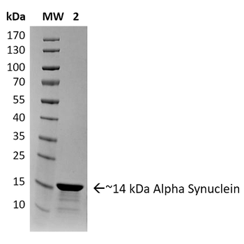
SDS-PAGE of ~14 kDa Human Recombinant Alpha Synuclein Protein Pre-formed Fibrils (Type 2). Lane 1: Molecular Weight Ladder (MW). Lane 2: Alpha Synuclein Protein Pre-formed Fibrils.
Primary rat hippocampal neurons show lewy body inclusion formation when treated with Type 1 Alpha Synuclein Pre-formed Fibrils at 4 µg/ml (D-F), but not when treated with Type 2 Alpha Synuclein Pre-formed Fibrils at 4 µg/ml (A-C). Tissue: Primary hippocampal neurons. Species: Sprague-Dawley rat. Fixation: 4% formaldehyde made from PFA. Primary Antibody: Mouse anti-pSer129 Antibody at 1:1000 24 hours at 4C, Secondary Antibody: FITC Goat Anti-Mouse (green) at 1:700 for 1 hours at RT (B, E). Counterstain: Hoechst (blue) nuclear stain at 1:4000 for 1 hour at RT (A, D). Localization: Lewy body inclusions. Magnification: 20x. C is A and B merged and F is D and E merged.
Type 1 alpha synuclein Pre-formed Fibrils seed the formation of new alpha synuclein fibrils from the pool of alpha synuclein monomers. Thioflavin T is a fluorescent dye that binds to beta sheet-rich structures, such as those in alpha synuclein fibrils. Upon binding, the emission spectrum of the dye experiences a red-shift and increased fluorescence intensity. Thioflavin T emission curves show increased fluorescence (correlated to alpha synuclein protein aggregation) over time when 10 µM of Type 1 alpha synuclein Pre-formed Fibrils is combined with 100 µM of alpha synuclein monomer, as compared to when 10 µM of Type 2 alpha synuclein Pre-formed Fibrils is combined with 100 µM of alpha synuclein monomer or 100 µM of alpha Synuclein monomer. Thioflavin T ex = 450 nm, em = 485 nm.
Toxicity results comparing Active Human Recombinant Alpha Synuclein Pre-formed Fibrils (Type 2) and Active Human Recombinant Alpha Synuclein Pre-formed Fibrils (Type 1). Data was graphed after live cell imaging results were obtained using the following procedure: After 8 days in vitro, primary rat mixed cortical neuron cells were washed with 1X PBS and treated with 500 µg/ml of Type 1 and Type 2 Alpha Synuclein Proteins for 20 hours at 37?C. Following treatements, cells were washed with 2X PBS and incubated with a staining solution (2.0 µM Cell Event + 2.5 µM Ethidium homodimer + 2.5 µg/ml Hoechst 33342 in sterile HBSS) for 30 minutes at 37?C. The addition of the Type 2 Alpha Synuclein Proteins resulted in a significant increase in cell death.
Evaluation of a-syn toxicity on primary mouse cortical neurons. Lactate dehydrogenase (LDH) is a soluble enzyme present in the cytosol that is released upon cell death. Toxicity was assessed with an LDH assay and displayed as % of vehicle control (VC). Data are presented as bar graphs and standard deviation.
Evaluation of a-syn toxicity on primary mouse cortical neurons. Mitochondrial dehydrogenase activity reduces yellow MTT to dark blue formazan crystals, a reaction catalyzed in living cells. Cell viability was assessed with an MTT assay and displayed as % of vehicle control (VC). Data are presented as bar graphs and standard deviation.
TEM of Type 2 Alpha Synuclein Pre-formed Fibrils (PFFs)
* Mehrwertsteuer und Versandkosten nicht enthalten. Irrtümer und Preisänderungen vorbehalten