Purified polyclonal antibody supplied in PBS with 0.09% (W/V) sodium azide. This antibody is purified through a protein A column, followed by peptide affinity purification.
Application Verdünnung:
IHC-P - 1:100-500, WB - 1:2000
All lanes: Anti-CD130 Antibody (C-term) at 1:2000 dilution. Lane 1: A549 whole cell lysate. Lane 2: HepG2 whole cell lysate. Lysates/proteins at 20 µg per lane. Secondary Goat Anti-Rabbit IgG, (H+L), Peroxidase conjugated at 1/10000 dilution. Predicted band size: 104 kDa. Blocking/Dilution buffer: 5% NFDM/TBST.
All lanes: Anti-CD130 Antibody (C-term) at 1:2000 dilution. Lane 1: HepG2 whole cell lysate. Lane 2: MDA-MB-231 whole cell lysate. Lane 3: SW480 whole cell lysate.Lysates/proteins at 20 µg per lane. Secondary Goat Anti-Rabbit IgG, (H+L), Peroxidase conjugated at 1/10000 dilution. Predicted band size: 104 kDa. Blocking/Dilution buffer: 5% NFDM/TBST.
All lanes: Anti-CD130 Antibody (C-term) at 1:2000 dilution. Lane 1: Hela whole cell lysate. Lane 2: MDA-MB-231 whole cell lysate. Lane 3: HepG2 whole cell lysate. Lane 4: SW480 whole cell lysate. Lane 5: A549 whole cell lysate.Lysates/proteins at 20 µg per lane. Secondary Goat Anti-Rabbit IgG, (H+L), Peroxidase conjugated at 1/10000 dilution. Predicted band size: 104 kDa. Blocking/Dilution buffer: 5% NFDM/TBST.
CD130 Antibody (C-term) western blot analysis in MDA-MB231 cell line lysates (35 ug/lane). This demonstrates the CD130 antibody detected the CD130 protein (arrow).
Anti-CD130 Antibody (C-term) at 1:2000 dilution + SW480 whole cell lysates.Lysates/proteins at 20 µg per lane. Secondary Goat Anti-Rabbit IgG, (H+L), Peroxidase conjugated at 1/10000 dilution. Predicted band size: 104 kDa. Blocking/Dilution buffer: 5% NFDM/TBST.
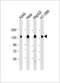
All lanes: Anti-CD130 Antibody (C-term) at 1:2000 dilution. Lane 1: A549 whole cell lysate. Lane 2: Hela whole cell lysate. Lane 3: HepG2 whole cell lysate. Lane 4: HT-1080 whole cell lysate.Lysates/proteins at 20 µg per lane. Secondary Goat Anti-Rabbit IgG, (H+L), Peroxidase conjugated at 1/10000 dilution. Predicted band size:104kDa. Blocking/Dilution buffer: 5% NFDM/TBST.
Staining CD130 in Human skeletal muscle tissue sections by Immunohistochemistry (IHC-P - paraformaldehyde-fixed, paraffin-embedded sections). Tissue was fixed with formaldehyde and blocked with 3% BSA for 0.5 hour at room temperature, antigen retrieval was by heat mediation with a citrate buffer (pH6). Samples were incubated with primary antibody (1/25) for 1 hours at 37C. A undiluted biotinylated goat polyvalent antibody was used as the secondary Antibody.
* Mehrwertsteuer und Versandkosten nicht enthalten. Irrtümer und Preisänderungen vorbehalten