Purified polyclonal antibody supplied in PBS with 0.09% (W/V) sodium azide. This antibody is purified through a protein A column, followed by peptide affinity purification.
Application Verdünnung:
FC - 1:25, WB - 1:1000, IHC-P - 1:100-500
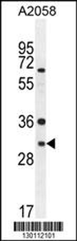
ARV1 Antibody (N-term) western blot analysis in A2058 cell line lysates (35 ug/lane). This demonstrates the ARV1 antibody detected the ARV1 protein (arrow).
ARV1 Antibody (N-term) western blot analysis in mouse Neuro-2a cell line lysates (35 ug/lane). This demonstrates the ARV1 antibody detected the ARV1 protein (arrow).
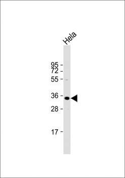
Anti-ARV1 Antibody (N-term) at 1:2000 dilution + Hela whole cell lysate.Lysates/proteins at 20 µg per lane. Secondary Goat Anti-Rabbit IgG, (H+L), Peroxidase conjugated at 1/10000 dilution. Predicted band size: 31 kDa. Blocking/Dilution buffer: 5% NFDM/TBST.
All lanes: Anti-ARV1 Antibody (N-term) at 1:2000 dilution. Lane 1: 293T/17 whole cell lysate. Lane 2: human kidney lysate. Lane 3: HL-60 whole cell lysate. Lane 4: K562 whole cell lysate.Lysates/proteins at 20 µg per lane. Secondary Goat Anti-Rabbit IgG, (H+L), Peroxidase conjugated at 1/10000 dilution. Predicted band size: 31 kDa. Blocking/Dilution buffer: 5% NFDM/TBST.
ARV1 antibody (N-term) immunohistochemistry analysis in formalin fixed and paraffin embedded human skeletal muscle followed by peroxidase conjugation of the secondary antibody and DAB staining. This data demonstrates the use of the ARV1 antibody (N-term) for immunohistochemistry. Clinical relevance has not been evaluated.
Overlay histogram showing A2058 cells (green line). The cells were fixed with 2% paraformaldehyde (10 min) and then permeabilized with 90% methanol for 10 min. The cells were then icubated in 2% bovine serum albumin to block non-specific protein-protein interactions followed by the antibody (1:25 dilution) for 60 min at 37C. The secondary antibody used was Goat-Anti-Rabbit IgG, DyLight 488 Conjugated Highly Cross-Adsorbed at 1/200 dilution for 40 min at 37C. Isotype control antibody (blue line) was rabbit IgG (1 µg/1x10 6 cells) used under the same conditions. Acquisition of > 10000 events was performed.
Overlay histogram showing A2058 cells (green line). The cells were fixed with 2% paraformaldehyde (10 min) and then permeabilized with 90% methanol for 10 min. The cells were then icubated in 2% bovine serum albumin to block non-specific protein-protein interactions followed by the antibody (1:25 dilution) for 60 min at 37C. The secondary antibody used was Goat-Anti-Rabbit IgG, DyLight 488 Conjugated Highly Cross-Adsorbed at 1/200 dilution for 40 min at 37C. Isotype control antibody (blue line) was rabbit IgG (1 µg/1x10 6 cells) used under the same conditions. Acquisition of > 10000 events was performed.
* Mehrwertsteuer und Versandkosten nicht enthalten. Irrtümer und Preisänderungen vorbehalten