Purified monoclonal antibody supplied in PBS with 0.09% (W/V) sodium azide. This antibody is purified through a protein G column, followed by dialysis against PBS.
Application Verdünnung:
WB - 1:200-2000, IF - 1:100, IHC-P - 1:100-500, FC - 1:10-50, IHC - 1:100
Fluorescent confocal image of SY5Y cells stained with SOX2 Antibody. SY5Y cells were fixed with 4% PFA (20 min), permeabilized with Triton X-100 (0.2%, 30 min). Cells were then incubated with SOX2 primary antibody (1:100, 2 h at room temperature). For secondary antibody, Alexa Fluor 488 conjugated donkey anti-mouse antibody (green) was used (1:1000, 1h).
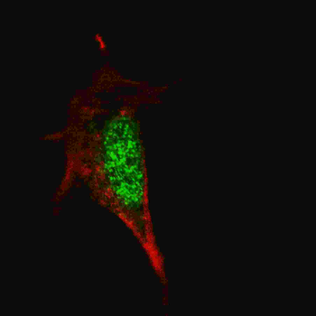
Fluorescent image of A549 cell stained with SOX2 Antibody. A549 cells were fixed with 4% PFA (20 min), permeabilized with Triton X-100 (0.1%, 10 min), then incubated with SOX2 primary antibody (1:25, 1 h at 37C. For secondary antibody, Alexa Fluor 488 conjugated donkey anti-mouse antibody (green) was used (1:400, 50 min at 37C.Cytoplasmic actin was counterstained with Alexa Fluor 555 (red) conjugated Phalloidin (7units/ml, 1 h at 37C.SOX2 immunoreactivity is localized to Nucleus significantly.
Western blot analysis of SOX2 (arrow) using mouse monoclonal SOX2 Antibody. 293 cell lysates (2 µg/lane) either nontransfected (Lane 1) or transiently transfected with the SOX2 gene (Lane 2)
Western blot analysis of SOX2 Antibody by SOX2 recombinant protein. SOX2 (arrow) was detected using the purified Mab.
Western blot analysis of lysate from NCCIT cell line, using SOX2 Antibody. diluted at 1:1000. A goat anti-mouse IgG H&L (HRP) at 1:3000 dilution was used as the secondary Antibody. Lysate at 20 µg.
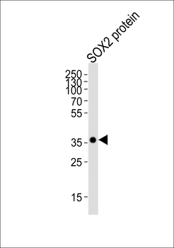
Western blot analysis of lysate from SOX2 protein, using SOX2 Antibody. diluted at 1:4000. A goat anti-mouse IgG H&L (HRP) at 1:3000 dilution was used as the secondary Antibody. Lysate at 20 µg.
Formalin-fixed and paraffin-embedded human lung carcinoma tissue reacted with SOX2 Antibody, which was peroxidase-conjugated to the secondary antibody, followed by DAB staining. This data demonstrates the use of this antibody for immunohistochemistry, clinical relevance has not been evaluated.
* Mehrwertsteuer und Versandkosten nicht enthalten. Irrtümer und Preisänderungen vorbehalten