Purified monoclonal antibody supplied in PBS with 0.09% (W/V) sodium azide. This antibody is purified through a protein G column, followed by dialysis against PBS.
Application Verdünnung:
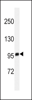
WB - 1:1000
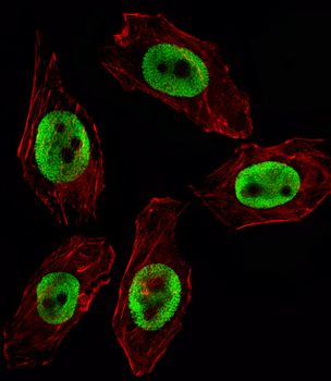
Fluorescent image of U251 cell stained with EZH2 Antibody. U251 cells were fixed with 4% PFA (20 min), permeabilized with Triton X-100 (0.1%, 10 min), then incubated with EZH2 primary antibody (1:25, 1 h at 37C. For secondary antibody, Alexa Fluor 488 conjugated donkey anti-mouse antibody (green) was used (1:400, 50 min at 37C. Cytoplasmic actin was counterstained with Alexa Fluor 555 (red) conjugated Phalloidin (7 units/ml, 1 h at 37C.EZH2 immunoreactivity is localized to Nucleus significantly.
Immunofluorescent analysis of 4% paraformaldehyde-fixed, 0.1% Triton X-100 permeabilized HeLa (human cervical epithelial adenocarcinoma cell line) cells labeling EZH2 at 1/25 dilution, followed by Dylight 488-conjugated goat anti-mouse IgG secondary antibody at 1/200 dilution (green). Immunofluorescence image showing nucleus staining on HeLa cell line. Cytoplasmic actin is detected with Dylight 554 Phalloidin at 1/100 dilution (red).
All lanes: Anti-EZH2 Antibody at 1:2000 dilution. Lane 1: Hela whole cell lysate. Lane 2: HL-60 whole cell lysate. Lane 3: T47D whole cell lysate.Lysates/proteins at 20 µg per lane. Secondary Goat Anti-mouse IgG, (H+L), Peroxidase conjugated at 1/10000 dilution. Predicted band size: 85 kDa. Blocking/Dilution buffer: 5% NFDM/TBST.
EZH2 Monoclonal Antibody immunohistochemistry analysis in formalin fixed and paraffin embedded human testis tissue followed by peroxidase conjugation of the secondary antibody and DAB staining. This data demonstrates the use of the EZH2 Monoclonal Antibody for immunohistochemistry. Clinical relevance has not been evaluated.
Staining EZH2 in human tonsil tissue sections by Immunohistochemistry (IHC-P - paraformaldehyde-fixed, paraffin-embedded sections). Tissue was fixed with formaldehyde and blocked with 3% BSA for 0.5 hour at room temperature, antigen retrieval was by heat mediation with a citrate buffer (pH6). Samples were incubated with primary antibody (1/25) for 1 hours at 37C. A undiluted biotinylated goat polyvalent antibody was used as the secondary Antibody.
Staining EZH2 in human colorectal carcinoma tissue sections by Immunohistochemistry (IHC-P - paraformaldehyde-fixed, paraffin-embedded sections). Tissue was fixed with formaldehyde and blocked with 3% BSA for 0.5 hour at room temperature, antigen retrieval was by heat mediation with a citrate buffer (pH6). Samples were incubated with primary antibody (1/25) for 1 hours at 37C. A undiluted biotinylated goat polyvalent antibody was used as the secondary Antibody.
Immunohistochemical analysis of paraffin-embedded Human tonsil section using Pink1. diluted at 1:50 dilution. A undiluted biotinylated goat polyvalent antibody was used as the secondary, followed by DAB staining.
* Mehrwertsteuer und Versandkosten nicht enthalten. Irrtümer und Preisänderungen vorbehalten