Purified monoclonal antibody supplied in PBS with 0.09% (W/V) sodium azide. This antibody is purified through a protein G column, followed by dialysis against PBS.
Application Verdünnung:
WB - 1:1000, IHC-P - 1:100-500, IF - 1:25
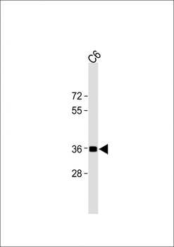
All lanes: Anti-GAPDH Antibody at 1:1000 dilution + C6 whole cell lysate. Lysates/proteins at 20 µg per lane. Secondary Goat Anti-mouse IgG, (H+L), Peroxidase conjugated at 1/15000 dilution.Observed band size: 36KDa. Blocking/Dilution buffer: 5% NFDM/TBST.
Fluorescent image of Hela cells stained with XAF1 GAPDH Antibody. diluted at 1:25 dilution. An Alexa Fluor 488-conjugated goat anti-mouse lgG at 1:400 dilution was used as the secondary antibody (green). Cytoplasmic actin was counterstained with Alexa Fluor 555 conjugated with Phalloidin (red).
All lanes: Anti-GAPDH Antibody at 1:1000 dilution. Lane 1: A431 whole cell lysates. Lane 2: C6 whole cell lysates. Lane 3: Hela whole cell lysates. Lane 4: HUVEC whole cell lysates.Lysates/proteins at 20 µg per lane. Secondary Goat Anti-mouse IgG, (H+L), Peroxidase conjugated at 1/10000 dilution. Predicted band size: 36 kDa. Blocking/Dilution buffer: 5% NFDM/TBST.
All lanes: Anti-GAPDH Antibody at 1:8000 dilution. Lane 1: Hela whole cell lysates. Lane 2: A549 whole cell lysates. Lane 3: COS-7 whole cell lysates. Lane 4: mouse brain lysates. Lane 5: C6 whole cell lysates. Lane 6: NIH/3T3 whole cell lysates. Lysates/proteins at 20 µg per lane. Secondary Goat Anti-mouse IgG, (H+L), Peroxidase conjugated at 1/10000 dilution. Predicted band size: 36 kDa. Blocking/Dilution buffer: 5% NFDM/TBST.
Western blot analysis of anti-GAPDH Monoclonal Antibody in CEM cell line lysates (35 µg/lane). GAPDH (arrow) was detected using the purified Mab.
Immunohistochemical analysis of paraffin-embedded H.kidney section using GAPDH Antibody. diluted at 1:25 dilution. A peroxidase-conjugated goat anti-mouse IgG at 1:400 dilution was used as the secondary antibody, followed by DAB staining.
Immunohistochemical analysis of paraffin-embedded H.stomach section using GAPDH Antibody. diluted at 1:25 dilution. A peroxidase-conjugated goat anti-mouse IgG at 1:400 dilution was used as the secondary antibody, followed by DAB staining.
* Mehrwertsteuer und Versandkosten nicht enthalten. Irrtümer und Preisänderungen vorbehalten