anti S100A9 antibody, anti S100 calcium binding protein A9 antibody, anti 60B8AG antibody, anti CAGB antibody, anti CFAG antibody, anti CGLB antibody, anti L1AG antibody, anti LIAG antibody, anti MAC387 antibody, anti MRP14 antibody, anti NIF antibody, anti P14 antibody, anti S100 calcium binding protein A9 (calgranulin B) antibody, anti S100 calcium-binding protein A9 antibody, anti S100 calcium-binding protein A9 (calgranulin B) antibody, anti calgranulin B antibody
Goat polyclonal antibody to S100A9
Klonalität:
Polyclonal
Molekulargewicht:
13.2
Puffer:
Supplied at 0.5 mg/ml in Tris saline, 0.02% sodium azide, pH 7.3 with 0.5% bovine serum albumin. Aliquot and store at -20C. Minimize freezing and thawing.
Sequenz:
DTNADKQLSFEEF
Target-Kategorie:
S100A9
Application Verdünnung:
ELISA: 1:8000, WB: 0.5-2 µg/ml, IHC-P: 2.5ug/ml
Anwendungsbeschreibung:
Application Notes: ELISA: Peptide ELISA: antibody detection limit dilution 1:8000.IHC: In paraffin embedded Human Lung shows nuclear staining in select cells, Recommended concentration, 2-4ug/ml.WB: Approx 16kDa band observed in Human Peripheral Blood Mononucleocytes lysates (calculated MW of 13.2kDa according to NP_002956.1). The observed molecular weight corresponds to earlier findings (~15kDa) in literature with different antibodies (McCormick et al, Biol Chem. 2005 Dec 16,280(50):41521-9., PMID: 16216873). Recommended concentration: 0.3-1 µg/ml
Negative Control showing staining of paraffin embedded Human Spleen, with no primary antibody.
Immunofluorescence analysis of paraformaldehyde fixed THP-1 cells immobilized on ShifixTM coverslip, permeabilized with 0.15% Triton. Primary incubation 1 hr (10 µg/mL) followed by Alexa Fluor 488 secondary antibody (2 µg/mL), showing nuclear and cytoplasmic staining. The nuclear stain is DAPI (blue). Negative control: Unimmunized goat IgG (10 µg/mL) followed by Alexa Fluor 488 secondary antibody (2 µg/mL).
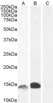
1 µg/mL staining of Human Bone Marrow (A) and (0.5 µg/mL) Gastrointestinal cancer (B) lysate and negative control HepG2 (C) lysate. 5 µg protein in RIPA buffer). Detected by chemiluminescence.
2.5 µg/mL staining of paraffin embedded Human Lung. Steamed antigen retrieval with citrate buffer pH 6, AP-staining.
Immunofluorescence analysis of paraformaldehyde fixed MCF7 cells, permeabilized with 0.15% Triton. Primary incubation 1 hr (10 µg/mL) followed by Alexa Fluor 488 secondary antibody (2 µg/mL), showing cytoplasmic and nuclear staining. The nuclear stain is DAPI (blue). Negative control: Unimmunized goat IgG (10 µg/mL) followed by Alexa Fluor 488 secondary antibody (2 µg/mL).
Immunofluorescence analysis of paraformaldehyde fixed U2OS cells, permeabilized with 0.15% Triton. Primary incubation 1 hr (10 µg/mL) followed by Alexa Fluor 488 secondary antibody (2 µg/mL), showing nuclear and cytoplasmic staining. The nuclear stain is DAPI (blue). Negative control: Unimmunized goat IgG (10 µg/mL) followed by Alexa Fluor 488 secondary antibody (2 µg/mL).
Flow cytometric analysis of paraformaldehyde fixed MCF7 cells (blue line), permeabilized with 0.5% Triton. Primary incubation 1 hr (10 µg/mL) followed by Alexa Fluor 488 secondary antibody (1 µg/mL). IgG control: Unimmunized goat IgG (black line) followed by Alexa Fluor 488 secondary antibody.
7 µg/mL staining of paraffin embedded Human Spleen. Heat induced antigen retrieval with citrate buffer pH 6, HRP-staining.
* Mehrwertsteuer und Versandkosten nicht enthalten. Irrtümer und Preisänderungen vorbehalten