E.coli-derived human Peroxiredoxin 3 recombinant protein (Position: T110-Q256). Human Peroxiredoxin 3 shares 93% amino acid (aa) sequence identity with both mouse and rat Peroxiredoxin 3.
Konjugation:
Unconjugated
Alternative Synonym:
Thioredoxin-dependent peroxide reductase, mitochondrial, 1.11.1.15, Antioxidant protein 1, AOP-1, HBC189, Peroxiredoxin III, Prx-III, Peroxiredoxin-3, Protein MER5 homolog, PRDX3, AOP1
Peroxiredoxin 3/PRDX3 Rabbit Polyclonal Antibody
Klonalität:
Polyclonal
Konzentration:
Adding 0.2 ml of distilled water will yield a concentration of 500 µg/ml.
Each vial contains antibody formulated with stabilizing components, 0.9 mg NaCl, 0.2 mg Na2HPO4, and 0.05 mg NaN3. *This antibody is supplied in a stabilized formulation. Compatibility with conjugation reactions depends on the chemistry of the conjugation
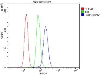
Flow Cytometry analysis of U937 cells using anti-Peroxiredoxin 3 antibody. Overlay histogram showing U937 cells (Blue line). To facilitate intracellular staining, cells were fixed with 4% paraformaldehyde and permeabilized with permeabilization buffer. The cells were blocked with 10% normal goat serum. And then incubated with rabbit anti-Peroxiredoxin 3 Antibody (1 µg/1x10 6 cells) for 30 min at 20C. DyLight488 conjugated goat anti-rabbit IgG (5-10 µg/1x10 6 cells) was used as secondary antibody for 30 minutes at 20C. Isotype control antibody (Green line) was rabbit IgG (1 µg/1x10 6) used under the same conditions. Unlabelled sample without incubation with primary antibody and secondary antibody (Red line) was used as a blank control.
WB analysis of Peroxiredoxin 3 using anti-Peroxiredoxin 3
IF analysis of Peroxiredoxin 3 using anti-Peroxiredoxin 3 antibody. Peroxiredoxin 3 was detected in immunocytochemical section of U20S cells. Enzyme antigen retrieval was performed using IHC enzyme antigen retrieval reagent for 15 mins. The cells were blocked with 10% goat serum. And then incubated with 2 µg/mL rabbit anti-Peroxiredoxin 3 Antibody overnight at 4C. DyLight594 Conjugated Goat Anti-Rabbit IgG was used as secondary antibody at 1:100 dilution and incubated for 30 minutes at 37C. The section was counterstained with DAPI. Visualize using a fluorescence microscope and filter sets appropriate for the label used.
IHC analysis of Peroxiredoxin 3 using anti-Peroxiredoxin 3 antibody. Peroxiredoxin 3 was detected in immunocytochemical section of Hela cell. Enzyme antigen retrieval was performed using IHC enzyme antigen retrieval reagent for 15 mins. The cells were blocked with 10% goat serum. And then incubated with 1 µg/ml rabbit anti-Peroxiredoxin 3 Antibody overnight at 4C. Biotinylated goat anti-rabbit IgG was used as secondary antibody and incubated for 30 minutes at 37C. The section was developed using Strepavidin-Biotin-Complex (SABC) with DAB as the chromogen.
IHC analysis of Peroxiredoxin 3 using anti-Peroxiredoxin 3 antibody. Peroxiredoxin 3 was detected in immunocytochemical section of MCF-7 cell. Enzyme antigen retrieval was performed using IHC enzyme antigen retrieval reagent for 15 mins. The cells were blocked with 10% goat serum. And then incubated with 1 µg/ml rabbit anti-Peroxiredoxin 3 Antibody overnight at 4C. Biotinylated goat anti-rabbit IgG was used as secondary antibody and incubated for 30 minutes at 37C. The section was developed using Strepavidin-Biotin-Complex (SABC) with DAB as the chromogen.
IHC analysis of Peroxiredoxin 3 using anti-Peroxiredoxin 3 antibody. Peroxiredoxin 3 was detected in immunocytochemical section of SMMC-7721 cell. Enzyme antigen retrieval was performed using IHC enzyme antigen retrieval reagent for 15 mins. The cells were blocked with 10% goat serum. And then incubated with 1 µg/ml rabbit anti-Peroxiredoxin 3 Antibody overnight at 4C. Biotinylated goat anti-rabbit IgG was used as secondary antibody and incubated for 30 minutes at 37C. The section was developed using Strepavidin-Biotin-Complex (SABC) with DAB as the chromogen.
IHC analysis of Peroxiredoxin 3 using anti-Peroxiredoxin 3 antibody. Peroxiredoxin 3 was detected in paraffin-embedded section of Human Intestinal Cancer Tissue. Heat mediated antigen retrieval was performed in citrate buffer (pH6, epitope retrieval solution) for 20 mins. The tissue section was blocked with 10% goat serum. The tissue section was then incubated with 1 µg/ml rabbit anti-Peroxiredoxin 3 Antibody overnight at 4C. Biotinylated goat anti-rabbit IgG was used as secondary antibody and incubated for 30 minutes at 37C. The tissue section was developed using Strepavidin-Biotin-Complex (SABC) with DAB as the chromogen.
Western blot analysis of Peroxiredoxin 3 using anti-Peroxiredoxin 3 antibody. Electrophoresis was performed on a 5-20% SDS-PAGE gel at 70V (Stacking gel) / 90V (Resolving gel) for 2-3 hours. The sample well of each lane was loaded with 50 ug of sample under reducing conditions. Lane 1: R
* Mehrwertsteuer und Versandkosten nicht enthalten. Irrtümer und Preisänderungen vorbehalten