KLH-conjugated synthetic peptide encompassing a sequence within the N-term region of human RBP5. The exact sequence is proprietary.
Konjugation:
Unconjugated
Alternative Synonym:
Retinol-binding protein 5, Cellular retinol-binding protein III, CRBP-III, HRBPiso
The RBP5 Antibody is suitable for IF, IHC, WB. It is a Polyclonal, Unconjugated antibody which raised against KLH-conjugated synthetic peptide encompassing a sequence within the N-term region of human RBP5. The exact sequence is proprietary. Purification: The antibody was purified by immunogen affinity chromatography.
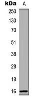
Western blot analysis of RBP5 expression in human liver (A) whole cell lysates. (Predicted band size: 15 kD, Observed band size: 16 kD)
Immunohistochemical analysis of RBP5 staining in human kidney formalin fixed paraffin embedded tissue section. The section was pre-treated using heat mediated antigen retrieval with sodium citrate buffer (pH 6.0). The section was then incubated with the antibody at room temperature and detected using an HRP conjugated compact polymer system. DAB was used as the chromogen. The section was then counterstained with haematoxylin and mounted with DPX.
Immunofluorescent analysis of RBP5 staining in LOVO cells. Formalin-fixed cells were permeabilized with 0.1% Triton X-100 in TBS for 5-10 minutes and blocked with 3% BSA-PBS for 30 minutes at room temperature. Cells were probed with the primary antibody in 3% BSA-PBS and incubated overnight at 4 C in a hidified chamber. Cells were washed with PBST and incubated with a DyLight 594-conjugated secondary antibody (red) in PBS at room temperature in the dark. DAPI was used to stain the cell nuclei (blue).
* Mehrwertsteuer und Versandkosten nicht enthalten. Irrtümer und Preisänderungen vorbehalten