A synthetic peptide corresponding to a sequence at the C-terminus of human HSD11B2, different from the related mouse sequence by five amino acids, and from the related rat sequence by three amino acids.
Konjugation:
Unconjugated
Alternative Synonym:
Corticosteroid 11-beta-dehydrogenase isozyme 2, 1.1.1.-, 11-beta-hydroxysteroid dehydrogenase type 2, 11-DH2, 11-beta-HSD2, 11-beta-hydroxysteroid dehydrogenase type II, 11-HSD type II, 11-beta-HSD type II, NAD-dependent 11-beta-hydroxysteroid dehydrogenase, 11-beta-HSD, Short chain dehydrogenase/reductase family 9C member 3, HSD11B2, HSD11K, SDR9C3
HSD11B2 Antibody
Klonalität:
Polyclonal
Konzentration:
Adding 0.2 ml of distilled water will yield a concentration of 500 µg/ml.
Application Notes: Western blot, 0.1-0.5µg/ml, Human, Mouse, Rat Immunohistochemistry (Paraffin-embedded Section), 0.5-1µg/ml, Human, Mouse, Rat Immunohistochemistry(Frozen Section), 2-5 µg/ml, Human, Mouse, Rat Immunofluorescence, 5 µg/ml, Human. Add 0.2ml of distilled water will yield a concentration of 500ug/ml
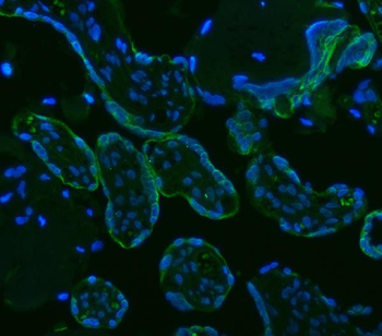
IF analysis of HSD11B2 using anti-HSD11B2 antibody. HSD11B2 was detected in a paraffin-embedded section of human placenta tissue. Heat mediated antigen retrieval was performed in EDTA buffer (pH8.0, epitope retrieval solution). The tissue section was blocked with 10% goat serum. The tissue section was then incubated with 5 µg/mL rabbit anti-HSD11B2 Antibody overnight at 4C. DyLight488 Conjugated Goat Anti-Rabbit IgG was used as secondary antibody at 1:500 dilution and incubated for 30 minutes at 37C. The section was counterstained with DAPI. Visualize using a fluorescence microscope and filter sets appropriate for the label used.
IHC analysis of HSD11B2 using anti-HSD11B2 antibody. HSD11B2 was detected in a frozen section of human placenta tissue. The tissue section was blocked with 10% goat serum. The tissue section was then incubated with 5 µg/ml rabbit anti-HSD11B2 Antibody overnight at 4C. Peroxidase Conjugated Goat Anti-rabbit IgG was used as secondary antibody and incubated for 30 minutes at 37C. The tissue section was developed using HRP Conjugated Rabbit IgG Super Vision Assay Kit with DAB as the chromogen.
IHC analysis of HSD11B2 using anti-HSD11B2 antibody. HSD11B2 was detected in a frozen section of mouse kidney tissue. The tissue section was blocked with 10% goat serum. The tissue section was then incubated with 2 µg/ml rabbit anti-HSD11B2 Antibody overnight at 4C. Peroxidase Conjugated Goat Anti-rabbit IgG was used as secondary antibody and incubated for 30 minutes at 37C. The tissue section was developed using HRP Conjugated Rabbit IgG Super Vision Assay Kit with DAB as the chromogen.
IHC analysis of HSD11B2 using anti-HSD11B2 antibody. HSD11B2 was detected in a frozen section of rat kidney tissue. The tissue section was blocked with 10% goat serum. The tissue section was then incubated with 2 µg/ml rabbit anti-HSD11B2 Antibody overnight at 4C. Peroxidase Conjugated Goat Anti-rabbit IgG was used as secondary antibody and incubated for 30 minutes at 37C. The tissue section was developed using HRP Conjugated Rabbit IgG Super Vision Assay Kit with DAB as the chromogen.
IHC analysis of HSD11B2 using anti-HSD11B2 antibody. HSD11B2 was detected in a paraffin-embedded section of Human Placenta tissue. Heat mediated antigen retrieval was performed in EDTA buffer (pH8.0, epitope retrieval solution). The tissue section was blocked with 10% goat serum. The tissue section was then incubated with 1 µg/ml rabbit anti-HSD11B2 Antibody overnight at 4C. Peroxidase Conjugated Goat Anti-rabbit IgG was used as secondary antibody and incubated for 30 minutes at 37C. The tissue section was developed using HRP Conjugated Rabbit IgG Super Vision Assay Kit with DAB as the chromogen.
IHC analysis of HSD11B2 using anti-HSD11B2 antibody. HSD11B2 was detected in a paraffin-embedded section of Mouse Pancreas tissue. Heat mediated antigen retrieval was performed in EDTA buffer (pH8.0, epitope retrieval solution). The tissue section was blocked with 10% goat serum. The tissue section was then incubated with 1 µg/ml rabbit anti-HSD11B2 Antibody overnight at 4C. Peroxidase Conjugated Goat Anti-rabbit IgG was used as secondary antibody and incubated for 30 minutes at 37C. The tissue section was developed using HRP Conjugated Rabbit IgG Super Vision Assay Kit with DAB as the chromogen.
IHC analysis of HSD11B2 using anti-HSD11B2 antibody. HSD11B2 was detected in a paraffin-embedded section of Rat Pancreas tissue. Heat mediated antigen retrieval was performed in EDTA buffer (pH8.0, epitope retrieval solution). The tissue section was blocked with 10% goat serum. The tissue section was then incubated with 1 µg/ml rabbit anti-HSD11B2 Antibody overnight at 4C. Peroxidase Conjugated Goat Anti-rabbit IgG was used as secondary antibody and incubated for 30 minutes at 37C. The tissue section was developed using HRP Conjugated Rabbit IgG Super Vision Assay Kit with DAB as the chromogen.
Western blot analysis of HSD11B2 using anti-HSD11B2 antib
* Mehrwertsteuer und Versandkosten nicht enthalten. Irrtümer und Preisänderungen vorbehalten