Each vial contains antibody formulated with stabilizing components, 0.9 mg NaCl, 0.2 mg Na2HPO4, and 0.05 mg NaN3. *This antibody is supplied in a stabilized formulation. Compatibility with conjugation reactions depends on the chemistry of the conjugation
Formulierung:
Lyophilized
Target-Kategorie:
Protein phosphatase 1 regulatory subunit 12A
Application Verdünnung:
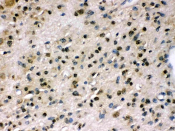
Western blot, 0.1-0.5µg/ml, Human, Mouse, Rat, Monkey Immunohistochemistry (Paraffin-embedded Section), 0.5-1µg/ml, Human Immunocytochemistry/Immunofluorescence, 2µg/ml, Human Flow Cytometry (Fixed), 1-3µg/1x10 6 cells, Human
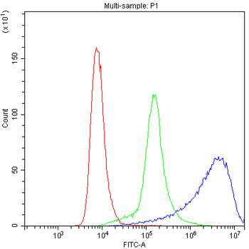
Flow Cytometry analysis of Hela cells using anti-PPP1R12A antibody. Overlay histogram showing Hela cells (Blue line). To facilitate intracellular staining, cells were fixed with 4% paraformaldehyde and permeabilized with permeabilization buffer. The cells were blocked with 10% normal goat serum. And then incubated with rabbit anti-PPP1R12A Antibody (1 µg/1x10 6 cells) for 30 min at 20C. DyLight488 conjugated goat anti-rabbit IgG (5-10 µg/1x10 6 cells) was used as secondary antibody for 30 minutes at 20C. Isotype control antibody (Green line) was rabbit IgG (1 µg/1x10 6) used under the same conditions. Unlabelled sample without incubation with primary antibody and secondary antibody (Red line) was used as a blank control.
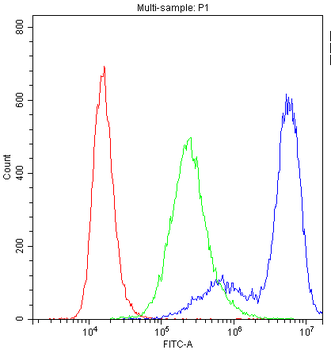
Flow Cytometry analysis of U251 cells using anti-PPP1R12A antibo
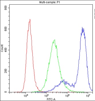
Flow Cytometry analysis of SiHa cells using anti-PPP1R12A antibody. Overlay histogram showing SiHa cells (Blue line). To facilitate intracellular staining, cells were fixed with 4% paraformaldehyde and permeabilized with permeabilization buffer. The cells were blocked with 10% normal goat serum. And then incubated with rabbit anti-PPP1R12A Antibody (1 µg/1x10 6 cells) for 30 min at 20C. DyLight488 conjugated goat anti-rabbit IgG (5-10 µg/1x10 6 cells) was used as secondary antibody for 30 minutes at 20C. Isotype control antibody (Green line) was rabbit IgG (1 µg/1x10 6) used under the same conditions. Unlabelled sample without incubation with primary antibody and secondary antibody (Red line) was used as a blank control.
Flow Cytometry analysis of U251 cells using anti-PPP1R12A antibody. Overlay histogram showing U251 cells (Blue line). To facilitate intracellular staining, cells were fixed with 4% paraformaldehyde and permeabilized with permeabilization buffer. The cells were blocked with 10% normal goat serum. And then incubated with rabbit anti-PPP1R12A Antibody (1 µg/1x10 6 cells) for 30 min at 20C. DyLight488 conjugated goat anti-rabbit IgG (5-10 µg/1x10 6 cells) was used as secondary antibody for 30 minutes at 20C. Isotype control antibody (Green line) was rabbit IgG (1 µg/1x10 6) used under the same conditions. Unlabelled sample without incubation with primary antibody and secondary antibody (Red line) was used as a blank control.
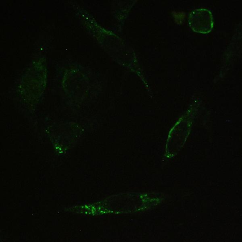
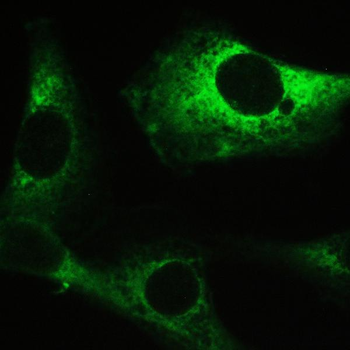
IHC analysis of PPP1R12A using anti-PPP1R12A antibody. PPP1R12A was detected in immunocytochemical section of SiHa cell. Enzyme antigen retrieval was performed using IHC enzyme antigen retrieval reagent for 15 mins. The cells were blocked with 10% goat serum. And then incubated with 2 µg/ml rabbit anti-PPP1R12A Antibody overnight at 4C. Biotin conjugated goat anti-rabbit IgG was used as secondary antibody and incubated for 30 minutes at 37C. The section was developed using DyLight488 Conjugated Avidin. Visualize using a fluorescence microscope and filter sets appropriate for the label used.
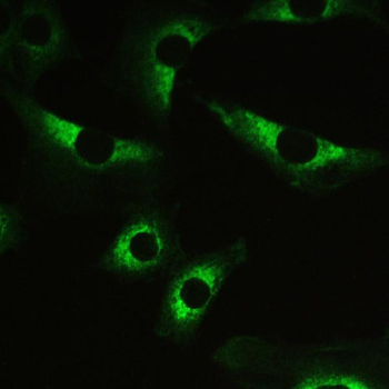
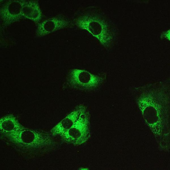
IHC analysis of PPP1R12A using anti-PPP1R12A antibody. PPP1R12A was detected in immunocytochemical section of U251 cell. Enzyme antigen retrieval was performed using IHC enzyme antigen retrieval reagent for 15 mins. The cells were blocked with 10% goat serum. And then incubated with 2 µg/ml rabbit anti-PPP1R12A Antibody overnight at 4C. Biotin conjugated goat anti-rabbit IgG was used as secondary antibody and incubated for 30 minutes at 37C. The section was developed using DyLight488 Conjugated Avidin. Visualize using a fluorescence microscope and filter sets appropriate for the label used.
IHC analysis of PPP1R12A using anti-PPP1R12A antibody. PPP1R12A was detected in immunocytochemical section of U251 cell. Enzyme antigen retrieval was performed using IHC enzyme antigen retrieval reagent for 15 mins. The cells were blocked with 10% goat serum. And then incubated with 2 µg/ml rabbit anti-PPP1R12A Antibody overnight at 4C. Biotin conjugated goat anti-rabbit IgG was used as secondary antibody and incubated for 30 minutes at 37C. The section was developed using DyLight488 Conjugated Avidin. Visualize using a fluorescence microscope and filter sets appropriate for the label
* Mehrwertsteuer und Versandkosten nicht enthalten. Irrtümer und Preisänderungen vorbehalten