Anti-AKT pS473 (MOUSE) Monoclonal Antibody was produced by repeated immunizations with a synthetic peptide corresponding to residues surrounding S473 of human AKT1 protein.
Anti-AKT pS473 (MOUSE) Monoclonal Antibody Biotin Conjugated was purified from concentrated tissue culture supernate by Protein A chromatography. This antibody is specific for human and mouse AKT protein phosphorylated at S473. A BLAST analysis was used to suggest cross-reactivity with AKT pS473 from human, mouse, rat and chimpanzee sources based on 100% homology with the immunizing sequence. Cross-reactivity with AKT from other sources has not been determined. Cross-reactivity with AKT2 and AKT3 has not been determined.
Application Notes: Biotin Conjugated Anti-AKT pS473 (MOUSE) Monoclonal Antibody Biotin Conjugated is tested for ELISA, immunohistochemistry, immunoprecipitation and western blotting. Expect a band approximately 56 kDa in size corresponding to phosphorylated AKT protein by western blotting in the appropriate cell lysate or extract. This phospho-specific monoclonal antibody reacts with human and mouse AKT pS473 and shows minimal reactivity by ELISA against the non-phosphorylated form of the immunizing peptide. Specific conditions for reactivity should be optimized by the end user. For immunohistochemistry use formalin-fixed paraffin-embedded sections. No pre-treatment of sample is required
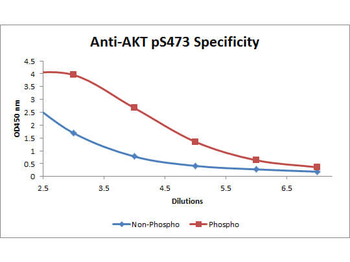
ELISA of Mouse anti-Akt phospho S473 Biotin Conjugated antibody. Antigen: BSA conjugates of Akt phospho S473 and AKT non-phospho S473. Coating amount: 0.1 µg per well. Primary antibody: Akt phospho S473 Biotin Conjugated antibody at 5 µg/ml. Dilution series: 3-fold. Mid-point concentration: 5 ng/mL Akt phospho S473 Biotin Conjugated antibody. Secondary antibody: Peroxidase streptavidin secondary antibody at 1:10000. Substrate: TMB.
Immunohistochemistry of mouse anti AKT phospho S473 biotin conjugated. Tissue: prostate at 40X (left) with negative control (right). Fixation: FFPE buffered formalin 10% conc. Antigen retrieval: Heat, Citrate pH6.2. Pressure Cooker. Primary antibody: AKT pS473 biotin at 20 µg/ml for 1 h at RT. Secondary antibody: Streptavidin Conj. HRP at 10 ug/ml. Localization: nuclear and occasionally cytoplasmic. Staining: antibody as precipitated red signal with a hematoxylin purple nuclear counterstain.
Immunohistochemistry of mouse Anti-AKT pS473 (MOUSE) Biotin Conjugated. Tissue: prostate at 40X. Fixation: FFPE buffered formalin 10% conc. Antigen retrieval: Heat, Citrate pH6.2. Pressure Cooker, left. (pH9 shown on right as negative control). Primary antibody: AKTsS473 biotin 20 µg/ml for 1 h at RT. Secondary antibody: Streptavidin Conj. HRP at 10 ug/ml. Localization: nuclear and occasionally cytoplasmic. Staining: antibody as precipitated red signal with a hematoxylin purple nuclear counterstain.
Western Blot of Mouse anti-Akt phospho S473 Biotin Conjugated antibody. Lane 1: GST tagged AKT1 active recombinant protein. Lane 2: none. Load: 25 ng per lane. Primary antibody: Akt phospho S473 Biotin Conjugated antibody at 1:1000 for overnight at 4C. Secondary antibody: HRP Streptavidin secondary antibody at 1:40000 for 30 min at RT. Block: orb348637 for 30 min at RT. Predicted/Observed size: 79 kDa, 79 kDa for Akt phospho S473. Other band(s): none
* Mehrwertsteuer und Versandkosten nicht enthalten. Irrtümer und Preisänderungen vorbehalten