Anti-AKT1 Antibody was produced in mice by repeated immunizations with a synthetic peptide corresponding to internal residues of human AKT1 protein followed by monoclonal development.
Anti-AKT1 antibody is directed against human AKT1. The antibody detects both unphosphorylated and phosphorylated forms of the protein. Anti-AKT1 antibody was purified from ascites by Protein A chromatography. Cross reactivity with AKT1 from other species has not been determined, however, the sequence of the immunogen shows 85% identity to mouse and 92% identity with rat, therefore, cross reactivity is expected.
Formulierung:
Lyophilized
Application Verdünnung:
ELISA: User Optimized, FC: User Optimized, IHC: User Optimized, IF: User Optimized, WB: User Optimized
Anwendungsbeschreibung:
Application Notes: Anti-AKT1 BIOTIN Antibody is suitable for Flow Cytometry, ELISA, immunohistochemistry, and western blotting. Expect a band approximately 56 kDa in size corresponding to AKT1 protein by western blotting in the appropriate cell lysate or extract. This monoclonal antibody reacts with human AKT. Specific conditions for reactivity should be optimized by the end user. For immunohistochemistry we recommend the use of fresh frozen tissues. Attempts at staining paraffin-embedded formalin fixed tissues were negative. No pre-treatment of sample is required
Flow Cytometry of Mouse anti-AKT1 antibody. Cells: LNCap Cells. Stimulation: none. Primary antibody: Allophycocyanin AKT1 antibody at 1.0 µg/ml for 20 min at 4C.
Plate was coated with monoclonal anti AKT1 antibody (capture antibody) followed by incubation with recombinant AKT1 (p/n orb346473), AKT2 (p/n orb346474), AKT3 (p/n orb346475) proteins. Binding was detected with biotinylated monoclonal anti-AKT pS473. The signal shows specificity of the monoclonal anti-AKT1 antibody to recombinant isoform AKT1 protein and not the isoform 2 and 3.
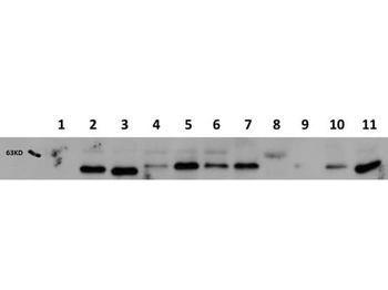
Western Blot of Mouse Anti-AKT1 antibody. Lane 1: AKT-1 Null. Lane 2: WT. Lane 3: MEF 1. Lane 4: A549. Lane 5: Calu-1. Lane 6: PC-3 (p/n orb692718). Lane 7: HepG2 (p/n orb348735). Lane 8: Jurkat (p/n orb348674). Lane 9: SKOV3. Lane 10: HEK293T. Lane 11: C2C12 (p/n orb348725). Load: 20 ug per lane. Primary antibody: AKT1 antibody at 1:1000 for overnight at 4C. Secondary antibody: Peroxidase mouse secondary antibody at 1:40000 for 30 min at RT. Block: orb348637 for 30 min at RT. Predicted/Observed size: 56 kDa for AKT1. Other band(s): none.
Western Blot of Mouse Anti-AKT1 antibody. Lane 1: GST Tagged recombinant AKT1. Lane 2: GST Tagged recombinant AKT2. Lane 3: GST Tagged recombinant AKT3. Load: 25 ng per lane. Primary antibody: AKT1 antibody at 1:1000 for overnight at 4C. Secondary antibody: Peroxidase mouse secondary antibody at 1:40000 for 30 min at RT. Block: orb348637 for 30 min at RT. Predicted/Observed size: 78 kDa for AKT1. Other band(s): none.
* Mehrwertsteuer und Versandkosten nicht enthalten. Irrtümer und Preisänderungen vorbehalten