Human Immunoglobulin Fab, F(ab), Fragment antigen-binding
Human IgG Fab Antibody
Konzentration:
2.1 mg/mL
Puffer:
Preservative: 0.01% (w/v) Sodium Azide. Stabilizer: None, Buffer: 0.02 M Potassium Phosphate, 0.15 M Sodium Chloride, pH 7.2
Quelle:
Human
Reinheit:
Human IgG Fab fragment was prepared from normal serum by a multi-step process which includes delipidation, salt fractionation and ion exchange chromatography followed by papain digestion and extensive dialysis against the buffer stated above. Human IgG Fab fragment assayed by immunoelectrophoresis resulted in a single precipitin arc against anti-Human Serum, anti- Human IgG and anti- Human IgG F(ab)2. No reaction was observed against anti- Human IgG F(c) or anti-Papain.
Formulierung:
Liquid (sterile filtered)
Anwendungsbeschreibung:
Biological Origin: Human. Application Notes: Human IgG Fab Fragment has been tested in SDS-Page and can be utilized as a control or standard reagent in Western Blotting and ELISA experiments
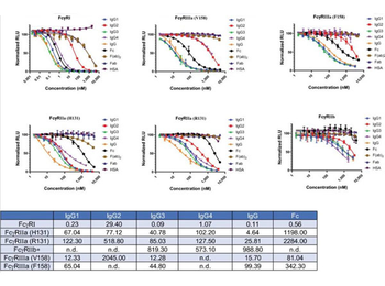
Dose dependent inhibition curves generated with six different FcgammaR assays. Four different set of samples were tested to show the specificity and subclass specific binding. Analytes tested are (1) human IgG subclasses IgG1, IgG2, IgG3, IgG4, (2) human IgG, (3) Fc, Fab, and F(ab)2 domains, and (4) human serum albumin (HSA). Data represent the mean standard error of triplicate experiments. IC50 (nM) values calculated from the inhibition curves are shown in the Table. IC50 values are in nM. *For FcgammaRIIb IC50 values are intended only for qualitative purposes as mentioned in the text. n.d. not determined.
SDS-Page of Human Fab. Lane 1: Human Fab - Non-Reduced. Lane 2: Human Fab - Reduced. Load: 1.0 ug per lane. Predicted/Observed size - Non-Reduced: 50 kDa, 50 kDa for Human Fab. Predicted/Observed size - Reduced: 25 kDa, 25 kDa for Human Fab. Other band(s): None.
SDS-PAGE results of Human IgG Fab Fragment. Lane 1: reduced Human IgG Fab Fragment (5 ug). Lane 2: reduced Human IgG Fab Fragment (1 ug). Lane 3: Opal Prestained Molecular Weight Ladder. Lane 4: non-reduced Human IgG Fab Fragment (1 ug). Lane 5: non-reduced Human IgG Fab Fragment (5 ug). 4-20% Lonza SDS-PAGE, Coomassie Stained, BioRad ChemiDoc Imaged.
SPR interaction analyses regarding the aptamer binding site in Protein A.Biacore X100 / sensor chip CAP / ligand: biotinylated Protein A with immobilization level of ~560 RU / two-step analyte binding without regeneration in between, (A-B) analyte 1 = sample 1: human IgG, IgG-Fc fragment, IgG-Fab fragment with a concentration of 1000 nM each, or buffer, (C-D) analyte 1 = sample 1: concentration series of human IgG-Fc in the range of 0-1000 nM, (A-D) analyte 2 = sample 2: 2000 nM 5-fluorescein-labeled aptamer PA2/8 or buffer. Double-referenced sensorgrams are shown (blank reference surface without Protein A, buffer injection). Binding of sample 1 followed by sample 2 is shown in (A) and (C) with alignment to injection start of sample 1. In (B) and (D) only binding of sample 2 with alignment to injection start of sample 2 is shown.
* Mehrwertsteuer und Versandkosten nicht enthalten. Irrtümer und Preisänderungen vorbehalten