A synthetic peptide corresponding to a sequence at the N-terminus of human MUC2, which shares 86.1% amino acid (aa) sequence identity with both mouse and rat MUC2.
Konjugation:
Unconjugated
Alternative Synonym:
MUC2, SMUC, Mucin-2, MUC-2, Intestinal mucin-2
MUC2 Rabbit Polyclonal Antibody
Klonalität:
Polyclonal
Konzentration:
Adding 0.2 ml of distilled water will yield a concentration of 500 µg/ml.
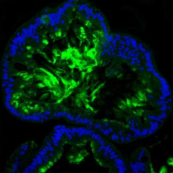
IF analysis of MUC2 using anti-MUC2 antibody. MUC2 was detected in paraffin-embedded section of human colon organoid tissue. Heat mediated antigen retrieval was performed in citrate buffer (pH6, epitope retrieval solution) for 20 mins. The tissue section was blocked with 10% goat serum. The tissue section was then incubated with 5 µg/mL rabbit anti-MUC2 Antibody overnight at 4C. DyLight488 Conjugated Goat Anti-Rabbit IgG was used as secondary antibody at 1:100 dilution and incubated for 30 minutes at 37C. The section was counterstained with DAPI. Visualize using a fluorescence microscope and filter sets appropriate for the label used.
IF analysis of MUC2 using anti-MUC2 antibody. MUC2 was detected in paraffin-embedded section of human ileum tissue. Heat mediated antigen retrieval was performed in citrate buffer (pH6, epitope retrieval solution) for 20 mins. The tissue section was blocked with 10% goat serum. The tissue section was then incubated with 5 µg/mL rabbit anti-MUC2 Antibody overnight at 4C. DyLight488 Conjugated Goat Anti-Rabbit IgG was used as secondary antibody at 1:100 dilution and incubated for 30 minutes at 37C. The section was counterstained with DAPI. Visualize using a fluorescence microscope and filter sets appropriate for the label used.
IF analysis of MUC2 using anti-MUC2 antibody. MUC2 was detected in paraffin-embedded section of human intestine cancer tissue. Heat mediated antigen retrieval was performed in citrate buffer (pH6, epitope retrieval solution) for 20 mins. The tissue section was blocked with 10% goat serum. The tissue section was then incubated with 5 µg/mL rabbit anti-MUC2 Antibody overnight at 4C. DyLight488 Conjugated Goat Anti-Rabbit IgG was used as secondary antibody at 1:100 dilution and incubated for 30 minutes at 37C. The section was counterstained with DAPI. Visualize using a fluorescence microscope and filter sets appropriate for the label used.
IF analysis of MUC2 using anti-MUC2 antibody. MUC2 was detected in paraffin-embedded section of mouse ileum organoid tissue. Heat mediated antigen retrieval was performed in citrate buffer (pH6, epitope retrieval solution) for 20 mins. The tissue section was blocked with 10% goat serum. The tissue section was then incubated with 5 µg/mL rabbit anti-MUC2 Antibody overnight at 4C. DyLight488 Conjugated Goat Anti-Rabbit IgG was used as secondary antibody at 1:100 dilution and incubated for 30 minutes at 37C. The section was counterstained with DAPI. Visualize using a fluorescence microscope and filter sets appropriate for the label used.
IF analysis of MUC2 using anti-MUC2 antibody. MUC2 was detected in paraffin-embedded section of mouse ileum tissue. Heat mediated antigen retrieval was performed in citrate buffer (pH6, epitope retrieval solution) for 20 mins. The tissue section was blocked with 10% goat serum. The tissue section was then incubated with 5 µg/mL rabbit anti-MUC2 Antibody overnight at 4C. DyLight488 Conjugated Goat Anti-Rabbit IgG was used as secondary antibody at 1:100 dilution and incubated for 30 minutes at 37C. The section was counterstained with DAPI. Visualize using a fluorescence microscope and filter sets appropriate for the label used.
IF analysis of MUC2 using anti-MUC2 antibody. MUC2 was detected in paraffin-embedded section of mouse intestine tissue. Heat mediated antigen retrieval was performed in citrate buffer (pH6, epitope retrieval solution) for 20 mins. The tissue section was blocked with 10% goat serum. The tissue section was then incubated with 5 µg/mL rabbit anti-MUC2 Antibody overnight at 4C. DyLight488 Conjugated Goat Anti-Rabbit IgG was used as secondary antibody at 1:100 dilution and incubated for 30 minutes at 37C. The section was counterstained with DAPI. Visualize using a fluorescence microscope and filter sets appropriate for the label used.
IHC analysis of MUC2 using anti-MUC2 antibody. MUC2 was detected in a paraffin-embedded section of human colon cancer tissue. Heat mediated anti
* Mehrwertsteuer und Versandkosten nicht enthalten. Irrtümer und Preisänderungen vorbehalten