E. coli-derived human Hsp20 recombinant protein (Position: H73-L123). Human Hsp20 shares 88.2% and 92.2% amino acid (aa) sequence identity with mouse and rat Hsp20, respectively.
Konjugation:
Unconjugated
Alternative Synonym:
Heat shock protein beta-6, HspB6, Heat shock 20 kDa-like protein p20, HSPB6
Hsp20/HSPB6 Rabbit Polyclonal Antibody
Klonalität:
Polyclonal
Konzentration:
Adding 0.2 ml of distilled water will yield a concentration of 500 µg/ml.
Western blot, 0.1-0.5µg/ml, Human, Mouse, Rat Immunohistochemistry (Paraffin-embedded Section), 0.5-1µg/ml, Human, Mouse, Rat Immunocytochemistry/Immunofluorescence, 2µg/ml, Human
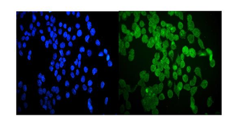
IF analysis of Hsp20 using anti-Hsp20 antibody. Hsp20 was detected in immunocytochemical section of A431 cells. Enzyme antigen retrieval was performed using IHC enzyme antigen retrieval reagent for 15 mins. The cells were blocked with 10% goat serum. And then incubated with 2 µg/mL rabbit anti-Hsp20 Antibody overnight at 4C. DyLight488 Conjugated Goat Anti-Rabbit IgG was used as secondary antibody at 1:100 dilution and incubated for 30 minutes at 37C. The section was counterstained with DAPI. Visualize using a fluorescence microscope and filter sets appropriate for the label used.
IF analysis of Hsp20 using anti-Hsp20 antibody. Hsp20 was detected in immunocytochem
IHC analysis of Hsp20 using anti-Hsp20 antibody. Hsp20 was detected in paraffin-embedded section of human mammary cancer tissue. Heat mediated antigen retrieval was performed in EDTA buffer (pH8.0, epitope retrieval solution). The tissue section was blocked with 10% goat serum. The tissue section was then incubated with 1 µg/ml rabbit anti-Hsp20 Antibody overnight at 4C. Biotinylated goat anti-rabbit IgG was used as secondary antibody and incubated for 30 minutes at 37C. The tissue section was developed using Strepavidin-Biotin-Complex (SABC) with DAB as the chromogen.
IHC analysis of Hsp20 using anti-Hsp20 antibody. Hsp20 was detected in paraffin-embedded section of human skeletal muscle tissue. Heat mediated antigen retrieval was performed in EDTA buffer (pH8.0, epitope retrieval solution). The tissue section was blocked with 10% goat serum. The tissue section was then incubated with 1 µg/ml rabbit anti-Hsp20 Antibody overnight at 4C. Biotinylated goat anti-rabbit IgG was used as secondary antibody and incubated for 30 minutes at 37C. The tissue section was developed using Strepavidin-Biotin-Complex (SABC) with DAB as the chromogen.
IHC analysis of Hsp20 using anti-Hsp20 antibody. Hsp20 was detected in paraffin-embedded section of mouse cardiac muscle tissue. Heat mediated antigen retrieval was performed in EDTA buffer (pH8.0, epitope retrieval solution). The tissue section was blocked with 10% goat serum. The tissue section was then incubated with 1 µg/ml rabbit anti-Hsp20 Antibody overnight at 4C. Biotinylated goat anti-rabbit IgG was used as secondary antibody and incubated for 30 minutes at 37C. The tissue section was developed using Strepavidin-Biotin-Complex (SABC) with DAB as the chromogen.
IHC analysis of Hsp20 using anti-Hsp20 antibody. Hsp20 was detected in paraffin-embedded section of mouse lung tissue. Heat mediated antigen retrieval was performed in EDTA buffer (pH8.0, epitope retrieval solution). The tissue section was blocked with 10% goat serum. The tissue section was then incubated with 1 µg/ml rabbit anti-Hsp20 Antibody overnight at 4C. Biotinylated goat anti-rabbit IgG was used as secondary antibody and incubated for 30 minutes at 37C. The tissue section was developed using Strepavidin-Biotin-Complex (SABC) with DAB as the chromogen.
IHC analysis of Hsp20 using anti-Hsp20 antibody. Hsp20 was detected in paraffin-embedded section of rat cardiac muscle tissue. Heat mediated antigen retrieval was performed in EDTA buffer (pH8.0, epitope retrieval solution). The tissue section was blocked with 10% goat serum. The tissue section was then incubated with 1 µg/ml rabbit anti-Hsp20 Antibody overnight at 4C. Biotinylated goat anti-rabbit IgG was used as secondary antibody and incubated for 30 minutes at 37C. The tissue section was developed using Strepavidin-Biotin-Complex (SABC) with DAB as the chromogen.
IHC analysis of Hsp20 using anti-Hsp20 antibody. Hsp20 was detected in paraffin-embedded section of rat lung tissue. Heat mediated antigen retrieval was performed in EDTA buffer (pH8.0, epitope retrieval solution). The tissue section was blocked with 10% goat serum. The tissue section was then incubated with 1 µg/ml rabbit anti-Hsp20 Antibody overnight at 4C. Biotinylated goat anti-rabbit IgG was used as secondary antibody and incubated for 30 minutes at 37C. The tissue section was developed using Strepavidin-Biotin-Compl
* Mehrwertsteuer und Versandkosten nicht enthalten. Irrtümer und Preisänderungen vorbehalten