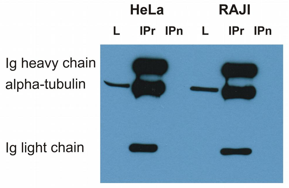
Immunoprecipitation of alpha-tubulin from HeLa and RAJI cell lysate by antibody TU-16 and its detection by antibody TU-01. IgM heavy chain (76-92 kDa) and IgM light chain (25-30 kDa) indicated. Mr of alpha tubulin is around 50 kDa.L = lysate, IPr = immunoprecipitate (reducing conditions), IPn = immunoprecipitate (non-reducing conditions).
Separation of HeLa cells stained using anti-alpha-Tubulin (TU-01) purified antibody (concentration in sample 3 µg/ml, GAM APC, red-filled) from HeLa cells unstained by primary antibody (GAM APC, black-dashed) in flow cytometry analysis (intracellular staining).
Immunocytochemistry staining of 3T3 mouse embryonal fibroblast cell line using anti-alpha-tubulin (TU-01, green) and anti-Vimentin (VI-01, cat. no. orb44570, red). Nucleus is stained with DAPI (blue).
Immunocytochemistry staining of HeLa human cervix carcinoma cell line using anti-alpha-tubulin (TU-01, red). Nucleus is stained with DAPI (blue).
Immunocytochemistry staining of 3T3 mouse embryonal fibroblast cell line using anti-alpha-tubulin (TU-01, green). Nucleus is stained with DAPI (blue).
Immunohistochemistry staining of human heart (paraffin sections) using anti-alpha-tubulin (TU-01).
Western blotting analysis of human alpha-tubulin using mouse monoclonal antibody TU-01 on lysates of various cell lines under reducing and non-reducing conditions. Nitrocellulose membrane was probed with 2 µg/ml of mouse anti-alpha-tubulin monoclonal antibody followed by IRDye800-conjugated anti-mouse secondary antibody. A specific band was detected for alpha-tubulin at approximately 54 kDa.
Use of anti-alpha-tubulin antibody TU-01 as a loading control (A) in an Western blotting experiment revealing the staining pattern of various cell lysates by a newly developed monoclonal antibody (B).
Anti-alpha-Tubulin Purified (TU-01) works in WB application under reducing conditions. Western blotting analysis was performed on whole cell extracts (RIPA lysis buffer) of JAR, JEG3, HTr-8/SVneo, and HeLa cell lines mixed and heated (100C, 5 min) with reducing (2-mercaptoethanol) or non-reducing SDS-loading buffer. Samples were resolved using 10% Tris-glycine SDS gel electrophoresis. Nitrocellulose membrane blot was probed simultaneously with mouse IgG1 monoclonal antibody TU-01 (1 µg/ml) and mouse IgM monoclonal antibody VI-01 detecting vimentin. Subclass-specific secondary antibodies IRDye 800CW Goat-anti-Mouse IgG (green) and IRDye 680RD Goat-anti-Mouse IgM (red) were used for multiplex fluorescent Western blot detection. Alpha-tubulin was detected at ~50 kDa and vimentin at ~55 kDa.
* Mehrwertsteuer und Versandkosten nicht enthalten. Irrtümer und Preisänderungen vorbehalten