
SMAD7 Rabbit Polyclonal Antibody, Unconjugated
Artikelnummer:
BYT-ORB500819
- Bilder (9)


| Artikelname: | SMAD7 Rabbit Polyclonal Antibody, Unconjugated |
| Artikelnummer: | BYT-ORB500819 |
| Hersteller Artikelnummer: | orb500819 |
| Alternativnummer: | BYT-ORB500819-50,BYT-ORB500819-100,BYT-ORB500819-200 |
| Hersteller: | Biorbyt |
| Wirt: | Rabbit |
| Kategorie: | Antikörper |
| Applikation: | FC, ICC, IF, IHC-Fr, IHC-P, WB |
| Spezies Reaktivität: | Human, Mouse, Rat |
| Immunogen: | KLH conjugated synthetic peptide derived from human Smad7 (1-100/426aa) |
| Konjugation: | Unconjugated |
| Alternative Synonym: | Madh7, SMAD7_HUMAN, SMAD7, MAD homolog 7, Mothers against DPP homolog 7, Mothers against decapentaplegic homolog 8 (MAD homolog 8 | Mothers against DPP homolog 8), SMAD family member 7 (SMAD 7 | Smad7 | hSMAD7), MADH8, SMAD7_MOUSE, SMAD family member 7 (SMAD 7 | Smad7), SMAD7_RAT, |
| SMAD7 Rabbit Polyclonal Antibody |
| Klonalität: | Polyclonal |
| Konzentration: | 1mg/ml |
| Molekulargewicht: | 46 kDa |
| UniProt: | O15105 |
| Puffer: | 0.01M TBS (pH7.4) with 1% rAlbumin, 0.02% Proclin300 and 50% Glycerol. |
| Formulierung: | Liquid |
| Target-Kategorie: | SMAD7 |
| Application Verdünnung: | WB=1:500-2000, IHC-P=1:100-500, IHC-F=1:100-500, ICC/IF=1:100-500, IF=1:100-500, Flow-Cyt=1ug/Test |
|
|
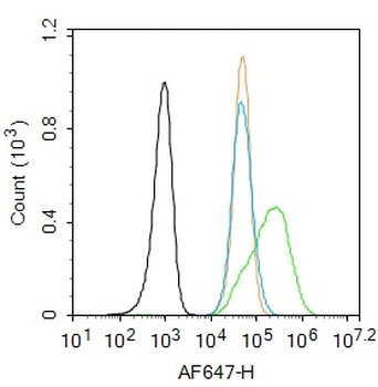
Blank control: SH-SY5Y. Primary Antibody (green line): Rabbit Anti-MADH7/Smad7 antibody (orb500819), dilution: 1 µg/10 6 cells, Isotype Control Antibody (orange line): Rabbit IgG. Secondary Antibody: Goat anti-rabbit IgG-AF647, dilution: 1 µg/Test. Protocol, The cells were fixed with 4% PFA (10 min at room temperature) and then permeabilized with 90% ice-cold methanol for 20 min at -20C. The cells were then incubated in 5% BSA to block non-specific protein-protein interactions for 30 min at room temperature. Cells stained with Primary Antibody for 30 min at room temperature. The secondary antibody used for 40 min at room temperature. Acquisition of 20000 events was performed. |
|
|
Paraformaldehyde-fixed, paraffin embedded (mouse kidney), Antigen retrieval by boiling in sodium citrate buffer (pH6.0) for 15 min, Block endogenous peroxidase by 3% hydrogen peroxide for 20 minutes, Blocking buffer (normal goat serum) at 37C for 30 min, Antibody incubation with (MADH7) Polyclonal Antibody, Unconjugated (orb500819) at 1:200 overnight at 4C, followed by operating according to SP Kit (Rabbit) instructionsand DAB staining. |
|
|
Paraformaldehyde-fixed, paraffin embedded (rat kidney), Antigen retrieval by boiling in sodium citrate buffer (pH6.0) for 15 min, Block endogenous peroxidase by 3% hydrogen peroxide for 20 minutes, Blocking buffer (normal goat serum) at 37C for 30 min, Antibody incubation with (MADH7) Polyclonal Antibody, Unconjugated (orb500819) at 1:200 overnight at 4C, followed by operating according to SP Kit (Rabbit) instructionsand DAB staining. |
|
|
Paraformaldehyde-fixed, paraffin embedded (rat lung), Antigen retrieval by boiling in sodium citrate buffer (pH6.0) for 15 min, Block endogenous peroxidase by 3% hydrogen peroxide for 20 minutes, Blocking buffer (normal goat serum) at 37C for 30 min, Antibody incubation with (Smad7) Polyclonal Antibody, Unconjugated (orb500819) at 1:600 overnight at 4C, followed by a conjugated secondary for 20 minutes and DAB staining. |
|
|
Paraformaldehyde-fixed, paraffin embedded (rat stomach), Antigen retrieval by boiling in sodium citrate buffer (pH6.0) for 15 min, Block endogenous peroxidase by 3% hydrogen peroxide for 20 minutes, Blocking buffer (normal goat serum) at 37C for 30 min, Antibody incubation with (MADH7) Polyclonal Antibody, Unconjugated (orb500819) at 1:200 overnight at 4C, followed by operating according to SP Kit (Rabbit) instructionsand DAB staining. |
|
|
Sample: Lane 1: Cerebrum (Mouse) Lysate at 40 ug, Lane 2: Cerebrum (Rat) Lysate at 40 ug, Lane 3: Stomach (Mouse) Lysate at 40 ug, Lane 4: Lung (Mouse) Lysate at 40 ug, Lane 5: Kidney (Mouse) Lysate at 40 ug, Primary: Anti-MADH7/Smad7 (orb500819) at 1/1000 dilution, Secondary: IRDye800CW Goat Anti-Rabbit IgG at 1/20000 dilution, Predicted band size: 50 kD, Observed band size: 50 kD. |
|
|
Sample: Lane 1: Stomach (Mouse) Lysate at 40 ug, Lane 2: Spleen (Mouse) Lysate at 40 ug, Lane 3: Lung (Mouse) Lysate at 40 ug, Primary: Anti-MADH7/Smad7 (orb500819) at 1/1000 dilution, Secondary: IRDye800CW Goat Anti-Rabbit IgG at 1/20000 dilution, Predicted band size: 50 kD, Observed band size: 50 kD. |
|
|
Sample: Lung (Mouse) Lysate at 30 ug, Placenta (Mouse) Lysate at 30 ug, Large intestine (Mouse) Lysate at 30 ug, Primary: Anti-MADH7/Smad7 (orb500819) at 1/300 dilution, Secondary: IRDye800CW Goat Anti-Rabbit IgG at 1/20000 dilution, Predicted band size: 46 kD, Observed band size: 50 kD. |
|
|
U-2OS cell, 4% Paraformaldehyde-fixed, Triton X-100 at room temperature for 20 min, Blocking buffer (normal goat serum) at 37C for 20 min, Antibody incubation with (MADH7/Smad7) polyclonal Antibody, Unconjugated (orb500819) 1:100, 90 minutes at 37C, followed by a conjugated Goat Anti-Rabbit IgG antibody at 37C for 90 minutes, DAPI (blue) was used to stain the cell nuclei. |
Produktgarantie und fachkundiger Support
