A synthetic peptide corresponding to a sequence at the N-terminus of human FH, which shares 100% and 97.8% amino acid (aa) sequence identity with mouse and rat FH, respectively.
Konjugation:
Unconjugated
Alternative Synonym:
Fumarate hydratase, mitochondrial, Fumarase, FH
Anti-FH Antibody (monoclonal, 9D8). Tested in IHC, WB applications. This antibody reacts with Human, Monkey, Mouse, Rat.
Klonalität:
Monoclonal
Konzentration:
Adding 0.2 ml of distilled water will yield a concentration of 500 µg/ml.
Western blot, 0.1-0.5µg/ml Immunohistochemistry (Paraffin-embedded Section), 0.5-1µg/ml
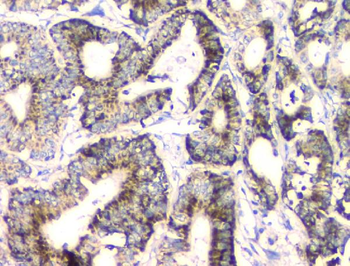
IHC analysis of FH using anti-FH antibody. FH was detected in paraffin-embedded section of human intestinal cancer tissue. Heat mediated antigen retrieval was performed in EDTA buffer (pH8.0, epitope retrieval solution). The tissue section was blocked with 10% goat serum. The tissue section was then incubated with 1 µg/ml mouse anti-FH Antibody overnight at 4C. Biotinylated goat anti-mouse IgG was used as secondary antibody and incubated for 30 minutes at 37C. The tissue section was developed using Strepavidin-Biotin-Complex (SABC) with DAB as the chromogen.
IHC analysis of FH using anti-FH antibody. FH was detected in paraffin-embedded section of human lung cancer tissue. Heat mediated antigen retrieval was performed in EDTA buffer (pH8.0, epitope retrieval solution). The tissue section was blocked with 10% goat serum. The tissue section was then incubated with 1 µg/ml mouse anti-FH Antibody overnight at 4C. Biotinylated goat anti-mouse IgG was used as secondary antibody and incubated for 30 minutes at 37C. The tissue section was developed using Strepavidin-Biotin-Complex (SABC) with DAB as the chromogen.
IHC analysis of FH using anti-FH antibody. FH was detected in paraffin-embedded section of rat liver tissue. Heat mediated antigen retrieval was performed in EDTA buffer (pH8.0, epitope retrieval solution). The tissue section was blocked with 10% goat serum. The tissue section was then incubated with 1 µg/ml mouse anti-FH Antibody overnight at 4C. Biotinylated goat anti-mouse IgG was used as secondary antibody and incubated for 30 minutes at 37C. The tissue section was developed using Strepavidin-Biotin-Complex (SABC) with DAB as the chromogen.
Western blot analysis of FH using anti-FH antibody. Electrophoresis was performed on a 5-20% SDS-PAGE gel at 70V (Stacking gel) / 90V (Resolving gel) for 2-3 hours. The sample well of each lane was loaded with 50 ug of sample under reducing conditions. Lane 1: K562 whole cell lysates, Lane 2: human placenta tissue lysates, Lane 3: COS-7 whole cell lysates, Lane 4: HL-60 whole cell lysates, Lane 5: Caco-2 whole cell lysates, Lane 6: U20S whole cell lysates, Lane 7: A549 whole cell lysates. After Electrophoresis, proteins were transferred to a Nitrocellulose membrane at 150mA for 50-90 minutes. Blocked the membrane with 5% Non-fat Milk/ TBS for 1.5 hour at RT. The membrane was incubated with mouse anti-FH antigen affinity purified monoclonal antibody at 0.5 µg/mL overnight at 4C, then washed with TBS-0.1% Tween 3 times with 5 minutes each and probed with a goat anti-mouse IgG-HRP secondary antibody at a dilution of 1:5000 for 1.5 hour at RT. The signal is developed using an Enhanced Chemiluminescent detection (ECL) kit with Tanon 5200 system. A specific band was detected for FH at approximately 48KD. The expected band size for FH is at 48KD.
Western blot analysis of FH using anti-FH antibody. Electrophoresis was performed on a 5-20% SDS-PAGE gel at 70V (Stacking gel) / 90V (Resolving gel) for 2-3 hours. The sample well of each lane was loaded with 50 ug of sample under reducing conditions. Lane 1: rat thymus tissue lysates, Lane 2: rat testicular tissue lysates, Lane 3: rat stomach tissue lysates, Lane 4: mouse testicular tissue lysates, Lane 5: mouse kidney tissue lysates, Lane 6: NIH3T3 whole cell lysates. After Electrophoresis, proteins were transferred to a Nitrocellulose membrane at 150mA for 50-90 minutes. Blocked the membrane with 5% Non-fat Milk/ TBS for 1.5 hour at RT. The membrane was incubated with mouse anti-FH antigen affinity purified monoclonal antibody at 0.5 µg/mL overnight at 4C, then washed with TBS-0.1% Tween 3 times with 5 minutes each and probed with a goat anti-mouse IgG-HRP secondary antibody at a dilution of 1:5000 for 1.5 hour at RT. The signal is developed using an Enhanced Chemiluminescent detection (ECL) kit with Tanon 5200 system. A specific band was detected for FH at approximately 48KD. The expected band size for FH is at 48KD.
* Mehrwertsteuer und Versandkosten nicht enthalten. Irrtümer und Preisänderungen vorbehalten